Copyright
©The Author(s) 2021.
World J Clin Cases. Nov 16, 2021; 9(32): 9990-9996
Published online Nov 16, 2021. doi: 10.12998/wjcc.v9.i32.9990
Published online Nov 16, 2021. doi: 10.12998/wjcc.v9.i32.9990
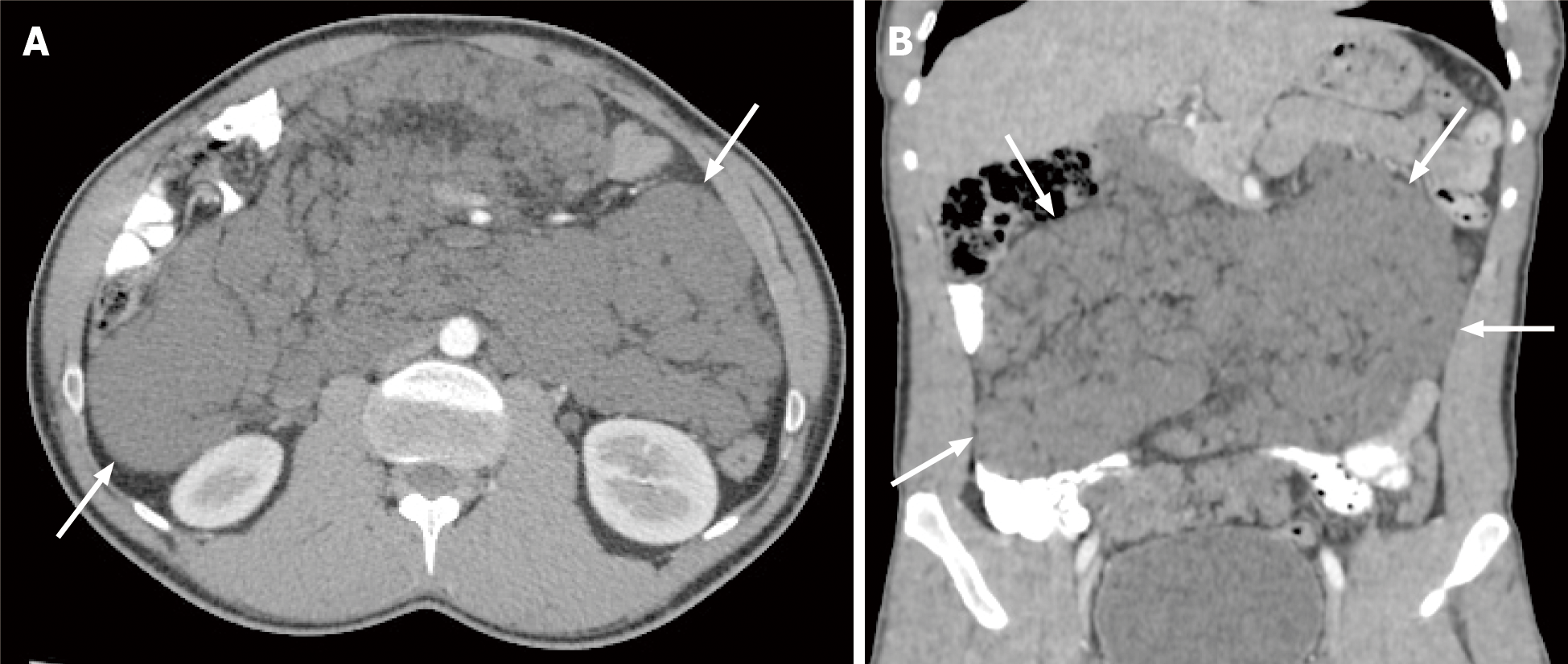
Figure 1 Computed tomography image of confluent peritoneal cystic lesions.
A: Post-contrast axial; B: Coronal computed tomography scan reveals numerous confluent peritoneal cystic lesions (white arrows) of variable sizes extensively involving the small bowel mesentery and responsible for peripheral displacement of the small bowel loops. No retro-peritoneal lesions are seen.
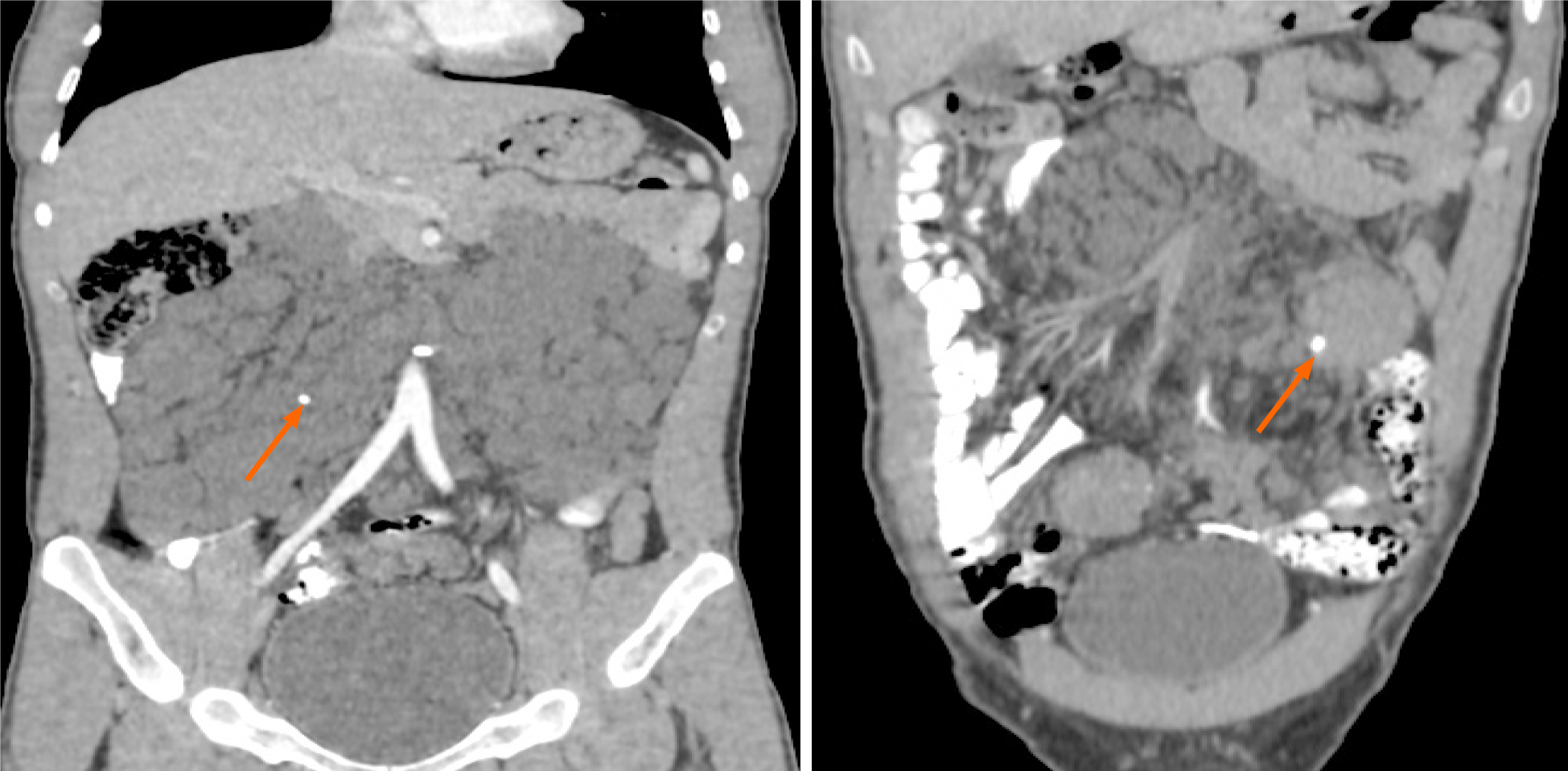
Figure 2 Post-contrast-enhanced coronal computed tomography scan revealed two small calcifications (orange arrows) within some of the lesions.
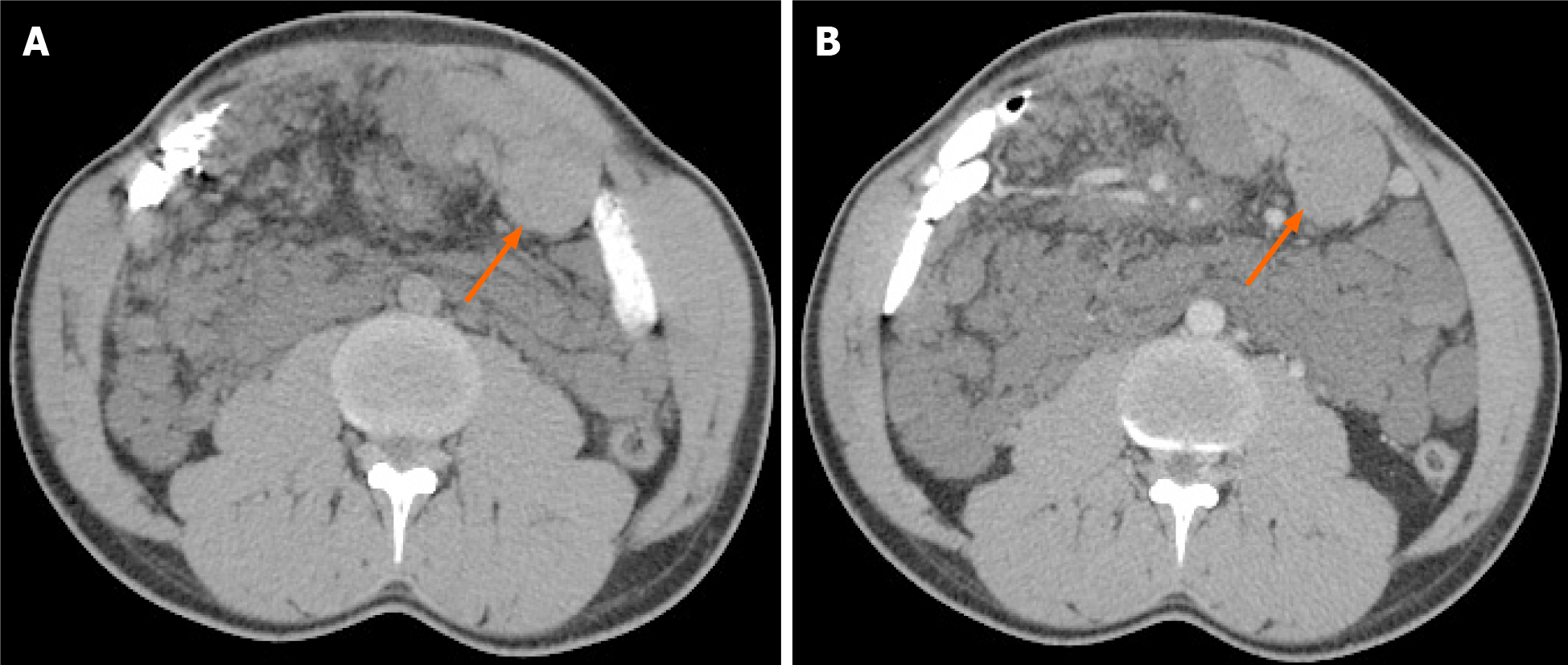
Figure 3 Computed tomography image of intra-cystic hemorrhage.
A: Non-enhanced axial computed tomography scan of the abdomen shows hemorrhage within one of the cystic lesions (orange arrow) located at the left lower quadrant with a spontaneous density of 53 Hounsfield units; B: Portal-venous phase axial contrast-enhanced image at the same level shows no internal enhancement within the lesion after administration of intravenous contrast.
- Citation: Alhasan AS, Daqqaq TS. Extensive abdominal lymphangiomatosis involving the small bowel mesentery: A case report. World J Clin Cases 2021; 9(32): 9990-9996
- URL: https://www.wjgnet.com/2307-8960/full/v9/i32/9990.htm
- DOI: https://dx.doi.org/10.12998/wjcc.v9.i32.9990


