Copyright
©The Author(s) 2021.
World J Clin Cases. Nov 16, 2021; 9(32): 9977-9981
Published online Nov 16, 2021. doi: 10.12998/wjcc.v9.i32.9977
Published online Nov 16, 2021. doi: 10.12998/wjcc.v9.i32.9977
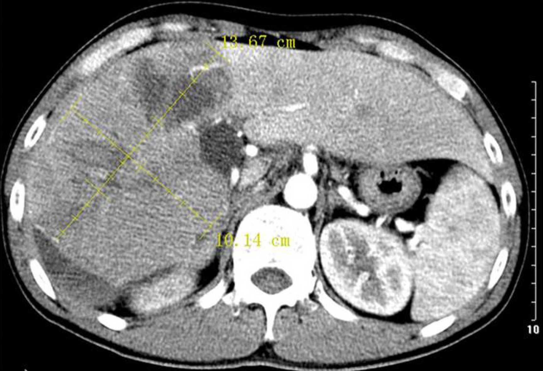
Figure 1 Computed tomography scan.
Large plaque-like lesion heterogeneously enhanced, including multiple cystic low-density lesions in the arterial phase with delayed portal washout, size 13.6 × 10.5 cm.
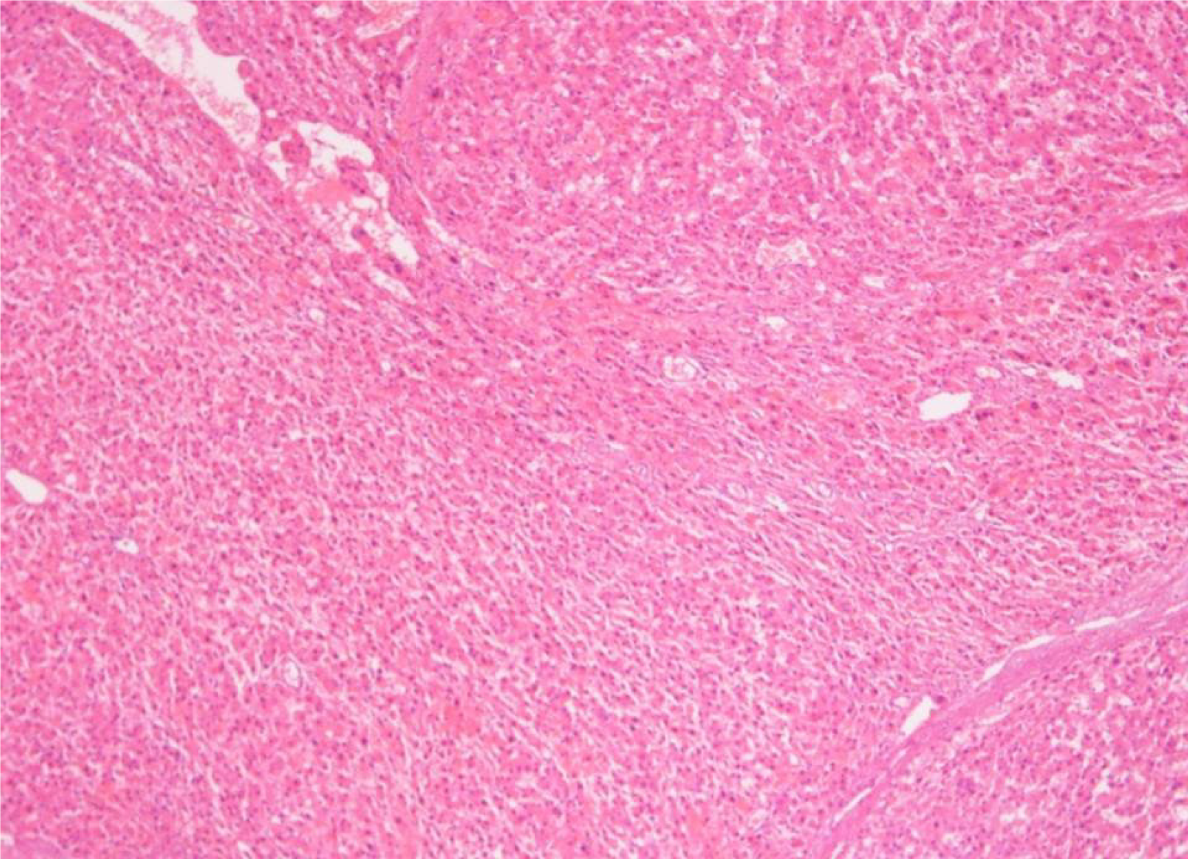
Figure 2 Postoperative pathology.
Hepatocytes with uniform size, arranged in clusters, with large dilated vessels and formation of fibrillar septum.
- Citation: Ren H, Gao YJ, Ma XM, Zhou ST. Large focal nodular hyperplasia is unresponsive to arterial embolization: A case report. World J Clin Cases 2021; 9(32): 9977-9981
- URL: https://www.wjgnet.com/2307-8960/full/v9/i32/9977.htm
- DOI: https://dx.doi.org/10.12998/wjcc.v9.i32.9977


