Copyright
©The Author(s) 2021.
World J Clin Cases. Nov 16, 2021; 9(32): 9948-9953
Published online Nov 16, 2021. doi: 10.12998/wjcc.v9.i32.9948
Published online Nov 16, 2021. doi: 10.12998/wjcc.v9.i32.9948
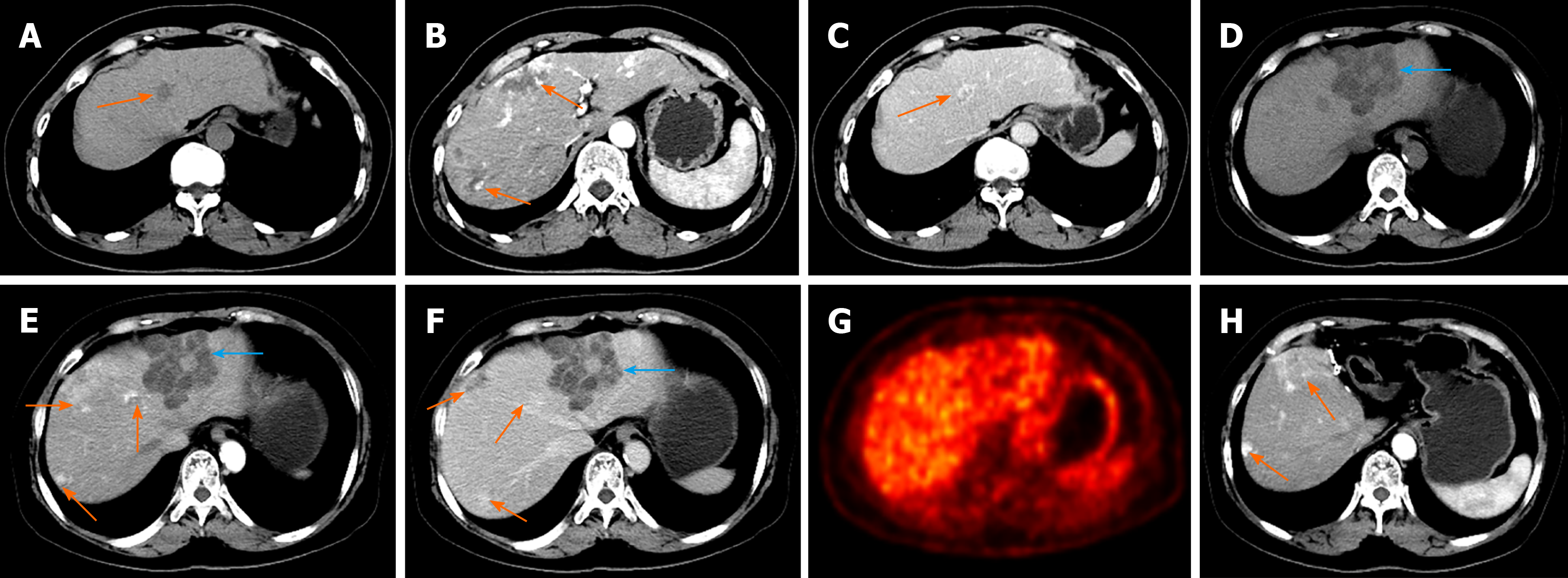
Figure 1 Contrast-enhanced computed tomography (CECT) and positron emission tomography/computed tomography (PET/CT) features of hepatic hemolymphangioma.
A–C: CECT images from 3 years ago revealing multiple hepatic hemangiomas (orange arrow) with hypointensity (A) and fast-in and slow-out enhancement patterns (B,C); D–F: CECT before partial hepatectomy presented a new cystic-solid lesion (blue arrow) with multiple internal divisions located in segment II of the liver (D), with delayed CECT enhancement characteristics (E, F); G: PET/CT scan found no significant uptake of 18F-fluorodeoxyglucose; H: At 1-year follow up, no obvious recurrent or residual lesion was identified by CT imaging.
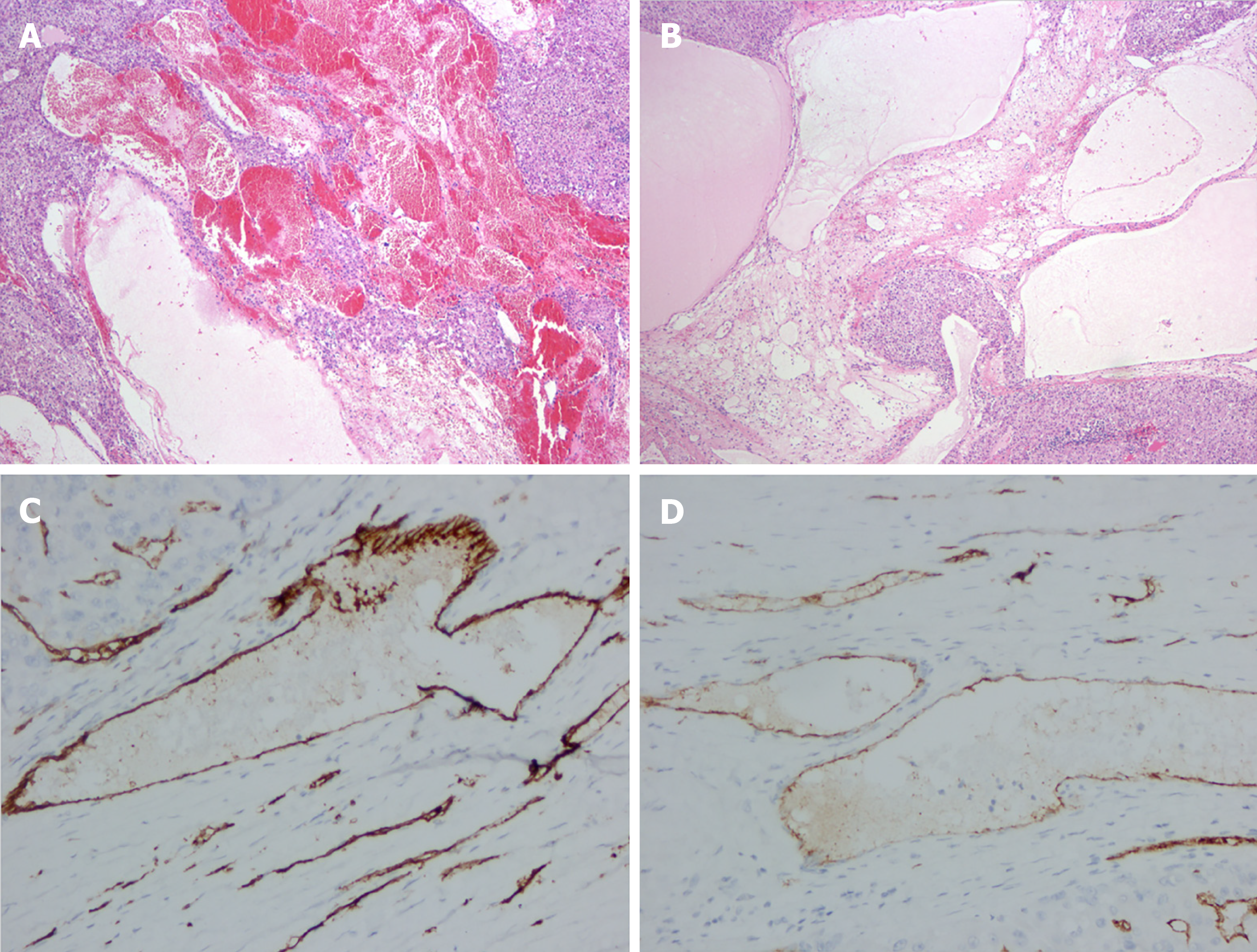
Figure 2 Pathological results of hepatic hemolymphangioma.
A, B: Microscopically, the tumor was mainly composed of lymphatic and blood vessels (40×); C, D: Immunohistochemical staining showed that CD34 and CD31 were positive (400×).
- Citation: Wang M, Liu HF, Zhang YZZ, Zou ZQ, Wu ZQ. Hemolymphangioma with multiple hemangiomas in liver of elderly woman with history of gynecological malignancy: A case report. World J Clin Cases 2021; 9(32): 9948-9953
- URL: https://www.wjgnet.com/2307-8960/full/v9/i32/9948.htm
- DOI: https://dx.doi.org/10.12998/wjcc.v9.i32.9948


