Copyright
©The Author(s) 2021.
World J Clin Cases. Nov 16, 2021; 9(32): 9926-9934
Published online Nov 16, 2021. doi: 10.12998/wjcc.v9.i32.9926
Published online Nov 16, 2021. doi: 10.12998/wjcc.v9.i32.9926
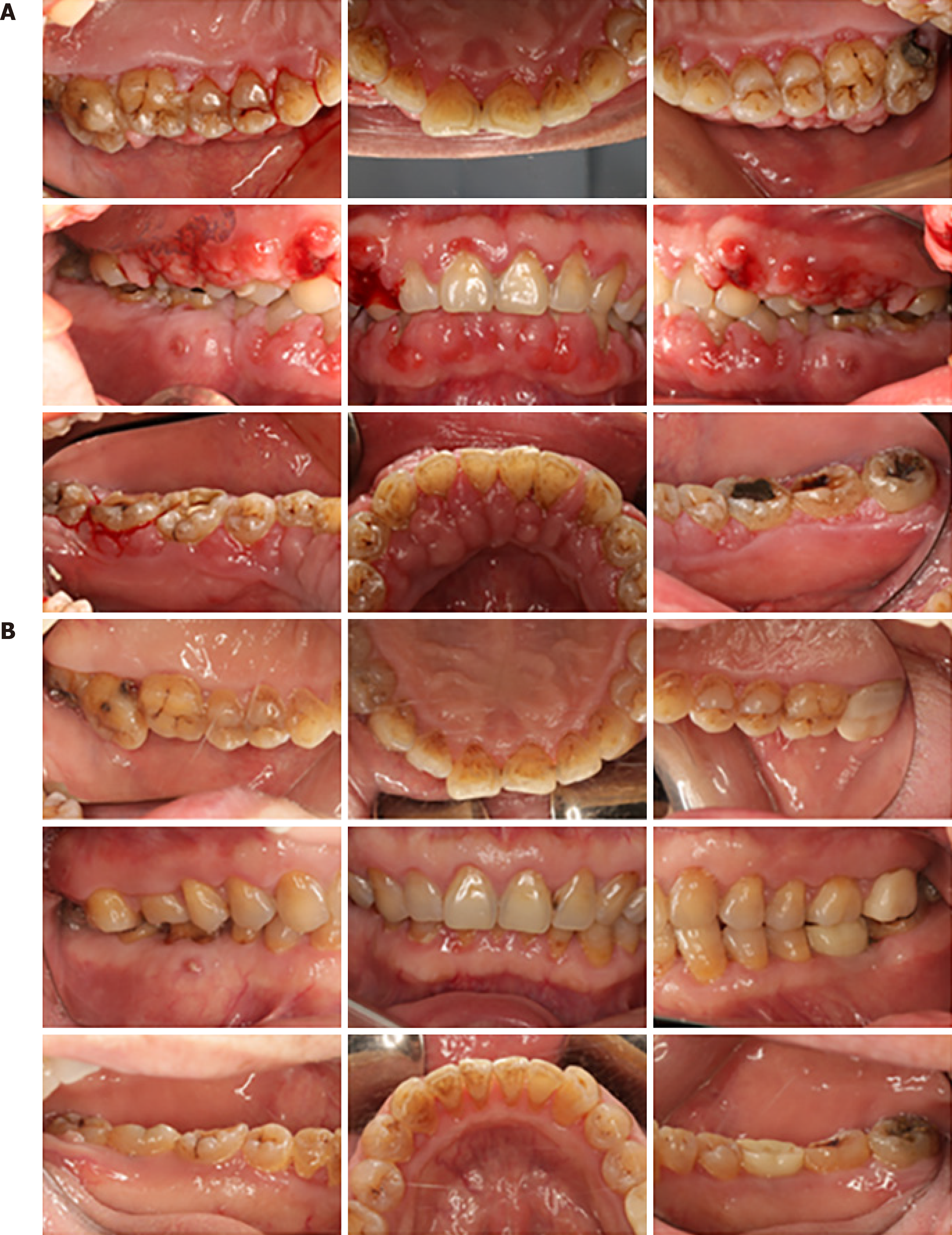
Figure 1 Imaging examination photos.
A: Initial clinical report on Patient 1 showing severe inflammatory and fibrotic GO with a spontaneous bleeding tendency. Fistula over tooth #46, the lower right first molar. A large silver–mercury filling in the crown of tooth #36. Tooth #27 had a large area of crown caries; B: Clinical re-evaluation after 2.5 years. The gingivae were less edematous and glazed. The fistula did not heal after root canal treatment in tooth #46. Later on, the patient requested removal of the affected tooth. The crown was repaired after root canal treatment of tooth #27. Tooth #36 was extracted due to fracture, and a dental implant was given.
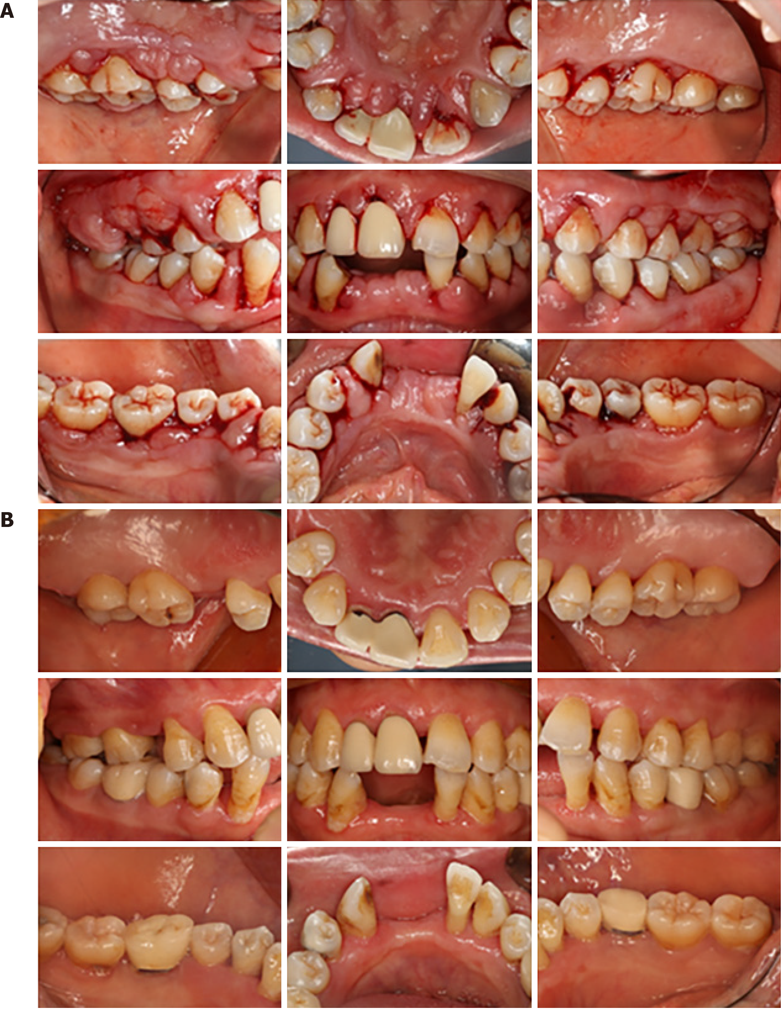
Figure 2 Imaging examination photos.
A: Initial clinical report of Patient 2 showing inflammatory and fibrotic GO with bleeding after periodontal probe. A 1–2-mm diastema between teeth #11 and #21, teeth#21 and #22, teeth #32 and #33, and teeth #43 and #44; B: Clinical re-evaluation 20 mo after periodontal surgery. Successful elimination of enlarged gingival tissue. The gingivae were less edematous and glazed. The crown was repaired after root canal treatment of teeth #35 and #46. The diastema was closed or reduced.
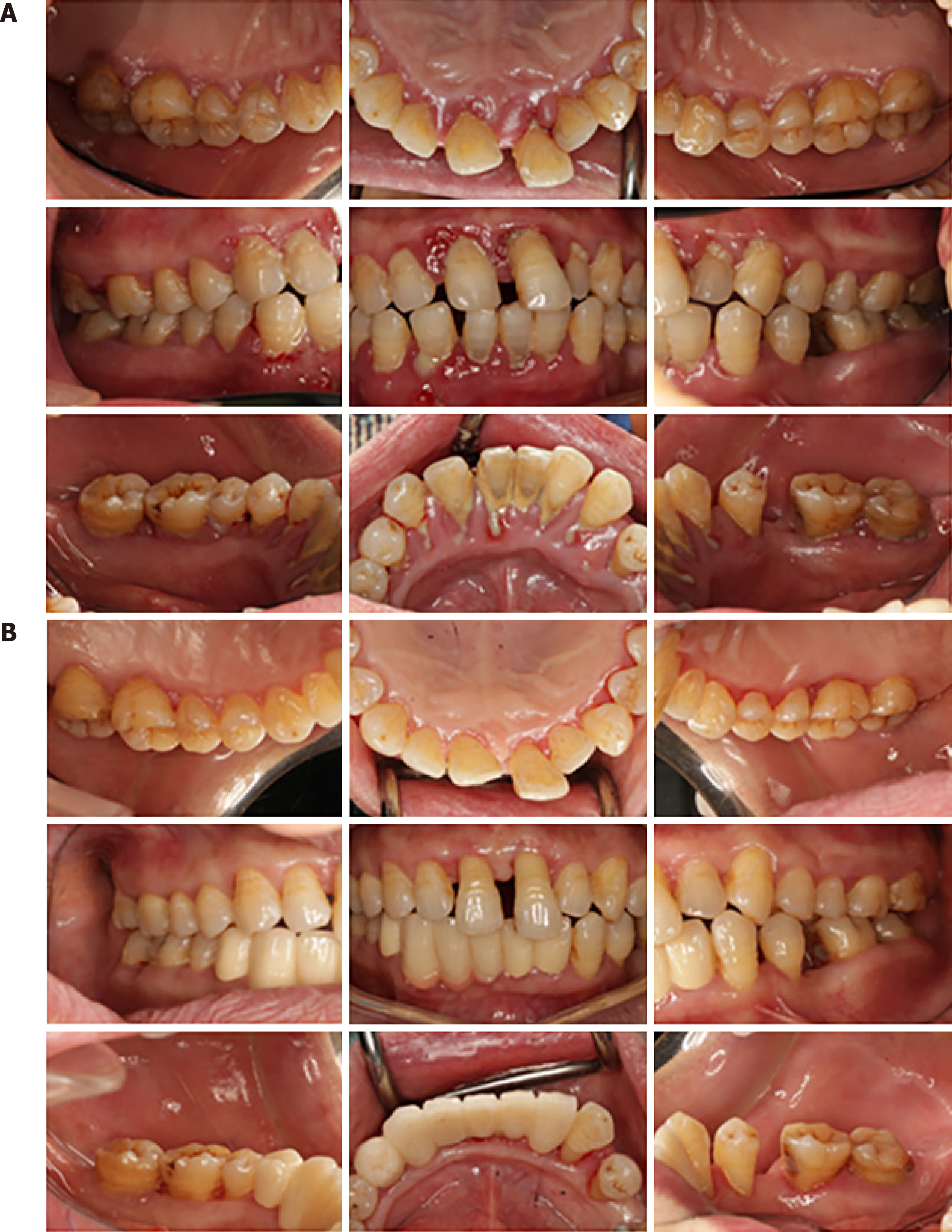
Figure 3 Imaging examination photos.
A: Initial clinical report of Patient 3 showing distinct inflammatory GO with generalized heavy sub- and supragingival calculus deposits. A 1–2.5-mm diastema between the upper and lower anterior teeth area; B: Clinical re-evaluation 14 mo after initial periodontal treatment. Successful elimination of enlarged gingival tissue. The diastema between the teeth has been closed or reduced. The crown was repaired after extraction of teeth with a poor prognosis.
Figure 4 Imaging examination photos.
A: Panoramic radiograph of Patient 1 showing mild-to-moderate generalized bone loss and severe furcation of tooth #46. Tooth #27 had a large area of crown caries with no pulp vitality. A large silver–mercury filling in the crown of tooth #36; B: Panoramic radiograph 1.5 years after treatment showing a mild-to-moderate generalized bone loss, with a mesial root of tooth #46 with complete bone loss. Tooth #36 was a dental implant.
Figure 5 Imaging examination photos.
A: The first panoramic radiograph of Patient 2 showing a generalized, severe bone loss; B: Panoramic radiograph of Patient 2 1 year after treatment showing no obvious alveolar bone loss compared to the initial examination. The alveolar bone density in the alveolar bone crest area was higher than that in the initial diagnosis. Teeth #28, #41, #42 and #31 had been removed after her first visit. Tooth #15 was extracted due to fracture.
Figure 6 Imaging examination photos.
A: Panoramic radiograph of Patient 3 showing generalized, severe bone loss. Teeth #31, #32, #41 and #42 were deemed hopeless. Tooth #43 showing a periapical shadow; B: Panoramic radiograph 14 mo after treatment showing no obvious alveolar bone loss compared to the initial examination; periodontal regeneration was found in some regions. Through the periodontal treatment combined with pulp treatment, regeneration was showed around the root of tooth #43.
- Citation: Fang L, Tan BC. Clinical presentation and management of drug-induced gingival overgrowth: A case series. World J Clin Cases 2021; 9(32): 9926-9934
- URL: https://www.wjgnet.com/2307-8960/full/v9/i32/9926.htm
- DOI: https://dx.doi.org/10.12998/wjcc.v9.i32.9926


