Copyright
©The Author(s) 2021.
World J Clin Cases. Nov 16, 2021; 9(32): 9917-9925
Published online Nov 16, 2021. doi: 10.12998/wjcc.v9.i32.9917
Published online Nov 16, 2021. doi: 10.12998/wjcc.v9.i32.9917
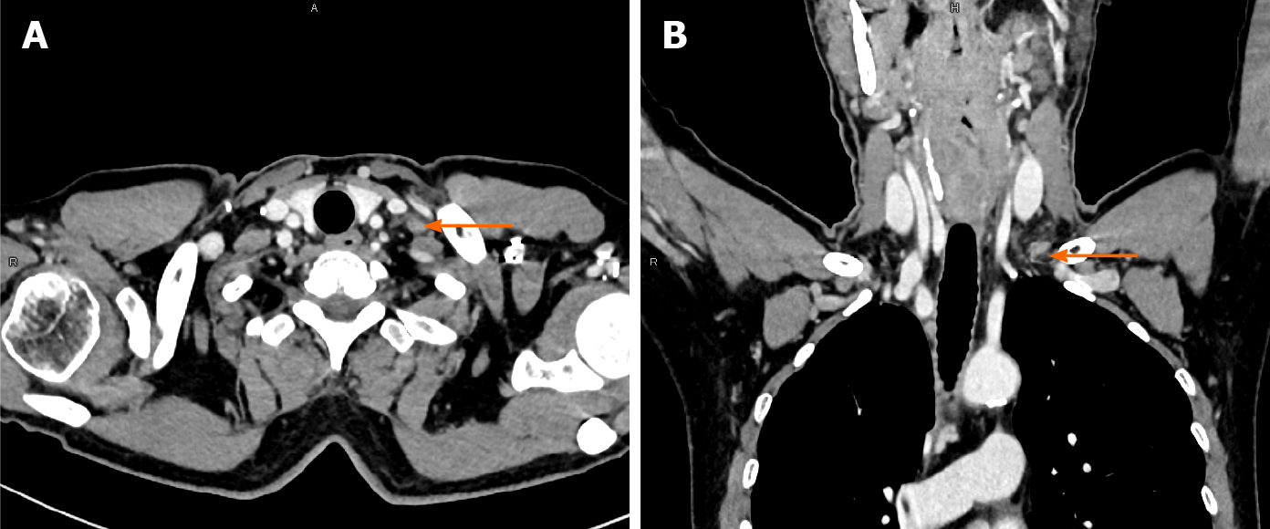
Figure 1 Contrast-enhanced computed tomography: The mass appeared to be mild enhancement.
A: Axial view; B: Coronal view.
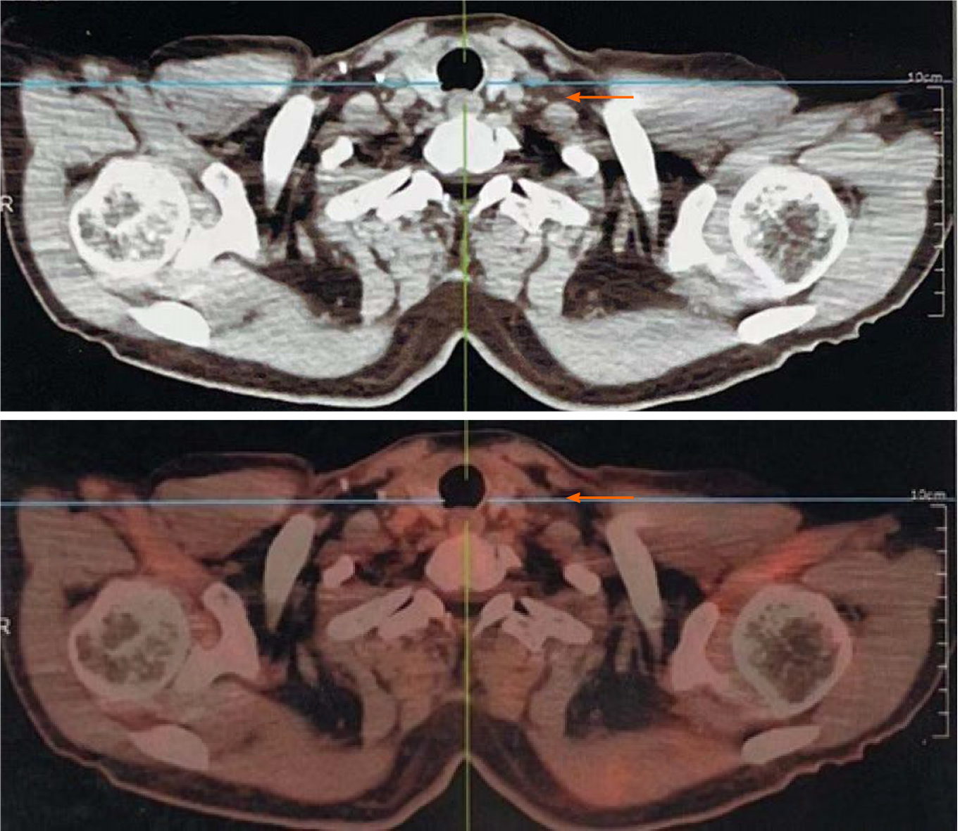
Figure 2 Positron emission tomography-computed tomography: No significant fluorodeoxyglucose uptake was observed in left supraclavicular region.
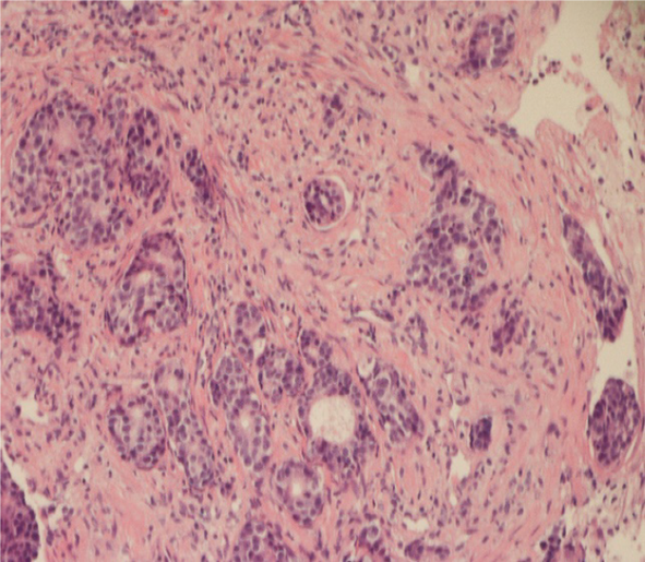
Figure 3 The left supraclavicular lymph node biopsy exhibited typical morphological findings of adenocarcinoma (hematoxylin & eosin staining × 200).
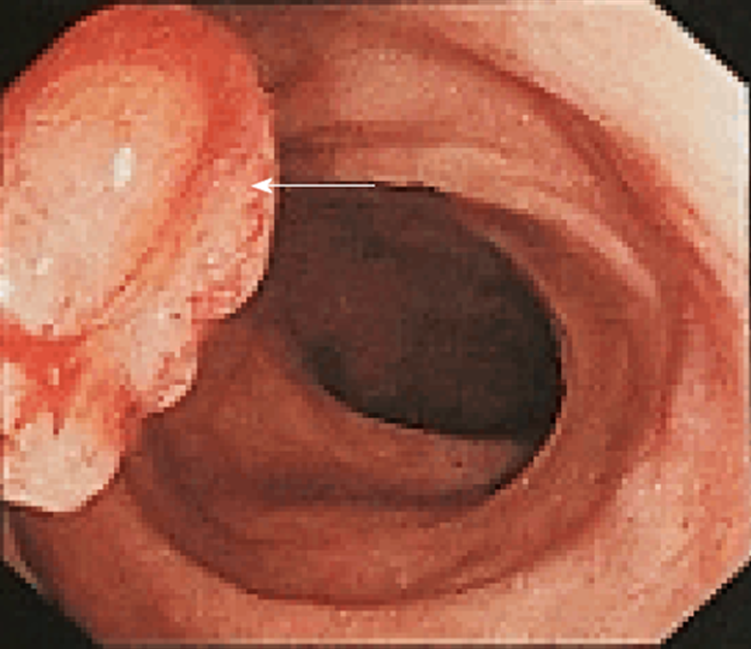
Figure 4 Colonoscopy examination: A pedunculated polyp measuring 2.
0 cm in diameter was observed in sigmoid colon.
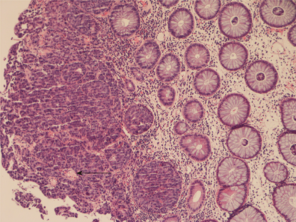
Figure 5 Pathological finding of the endoscopic biopsy (hematoxylin & eosin staining × 100): Carcinoma tissue invades the muscularis mucosa.
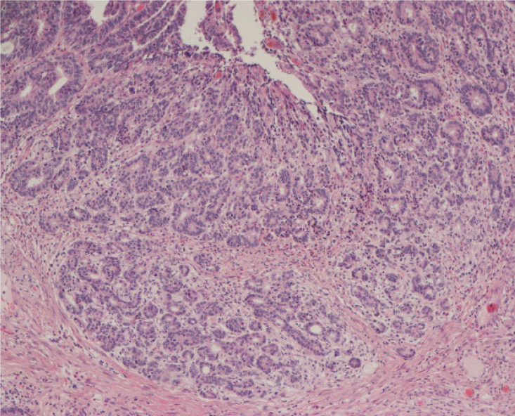
Figure 6 Pathological findings of sigmoid tumor (hematoxylin & eosin staining × 100): Moderately differentiated adenocarcinomas limited to mucous membranes.
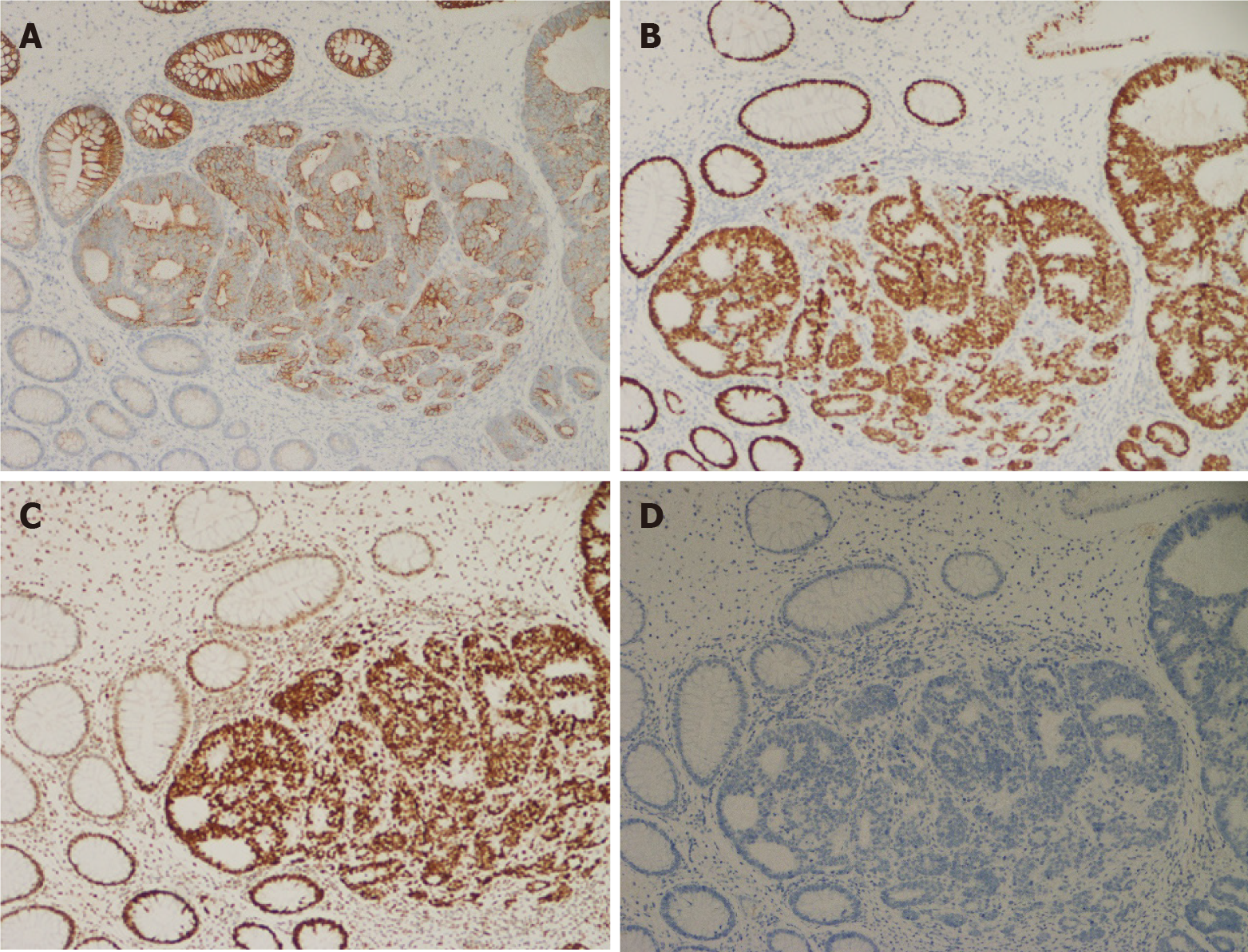
Figure 7 Immunohistochemical examination of sigmoid tumor (hematoxylin & eosin staining × 100).
A: CK20 is positive; B: MSH2 is positive; C: SATB2 is positive; D: CK7 is negative.
- Citation: Yang JQ, Shang L, Li LP, Jing HY, Dong KD, Jiao J, Ye CS, Ren HC, Xu QF, Huang P, Liu J. Isolated synchronous Virchow lymph node metastasis of sigmoid cancer: A case report. World J Clin Cases 2021; 9(32): 9917-9925
- URL: https://www.wjgnet.com/2307-8960/full/v9/i32/9917.htm
- DOI: https://dx.doi.org/10.12998/wjcc.v9.i32.9917


