Copyright
©The Author(s) 2021.
World J Clin Cases. Nov 16, 2021; 9(32): 9896-9902
Published online Nov 16, 2021. doi: 10.12998/wjcc.v9.i32.9896
Published online Nov 16, 2021. doi: 10.12998/wjcc.v9.i32.9896
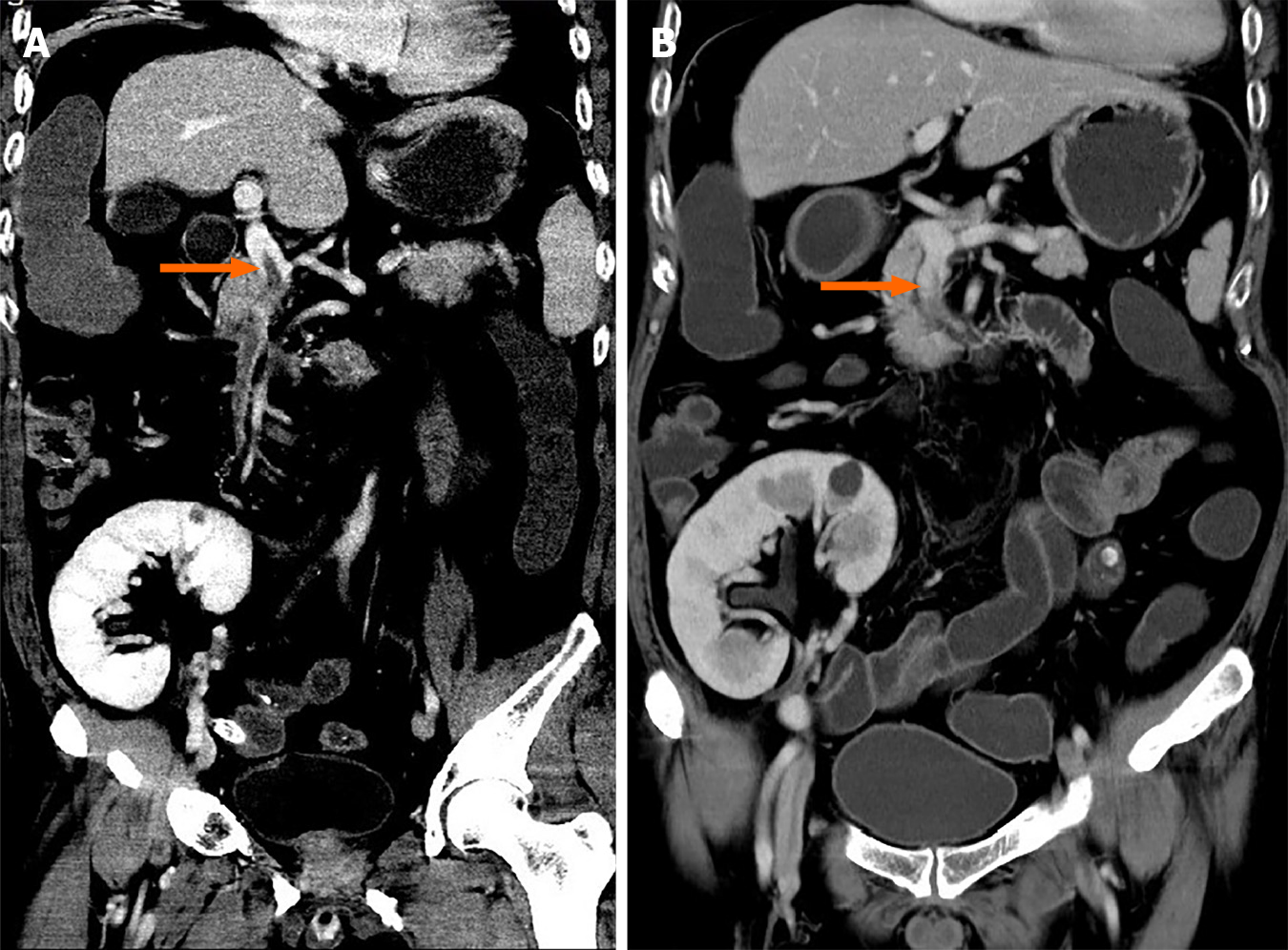
Figure 1 Whole abdomen enhanced computed tomography scan.
A: An acute mesenteric venous thrombosis (orange arrow); B: Recanalization after 2 mo of anticoagulation (orange arrow).
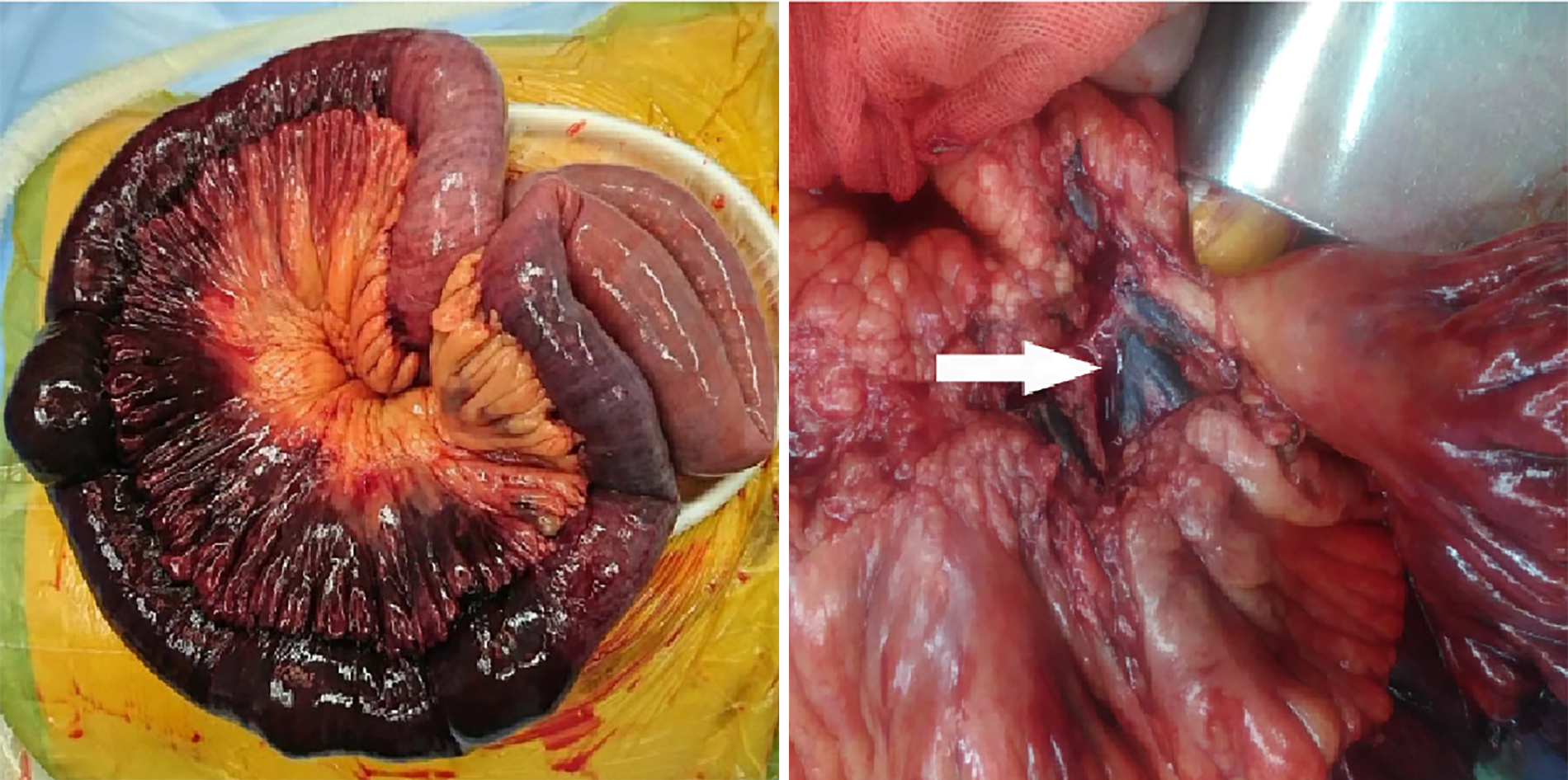
Figure 2
Intraoperative images of infarcted small bowel secondary to thrombosis of mesenteric vein (as indicated by white arrow).
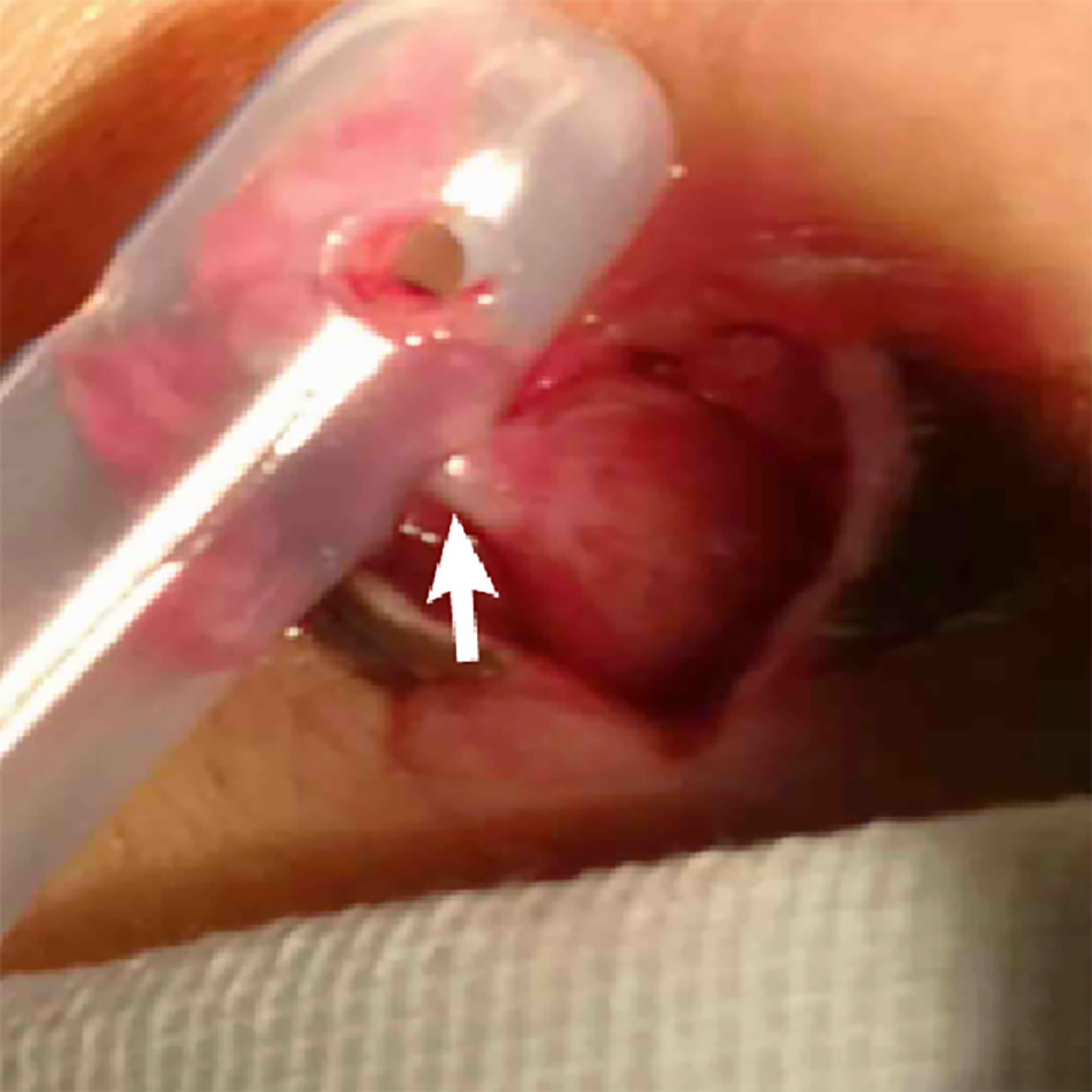
Figure 3
A rare hernia, partial intestinal wall (white arrow) incarcerated in the hole of drainage tube.
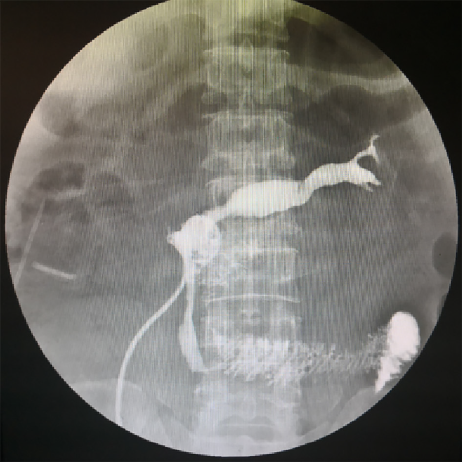
Figure 4
Fistula formation at the anastomotic site.
- Citation: Zhang P, Li XJ, Guo RM, Hu KP, Xu SL, Liu B, Wang QL. Idiopathic acute superior mesenteric venous thrombosis after renal transplantation: A case report. World J Clin Cases 2021; 9(32): 9896-9902
- URL: https://www.wjgnet.com/2307-8960/full/v9/i32/9896.htm
- DOI: https://dx.doi.org/10.12998/wjcc.v9.i32.9896


