Copyright
©The Author(s) 2021.
World J Clin Cases. Nov 16, 2021; 9(32): 9752-9761
Published online Nov 16, 2021. doi: 10.12998/wjcc.v9.i32.9752
Published online Nov 16, 2021. doi: 10.12998/wjcc.v9.i32.9752
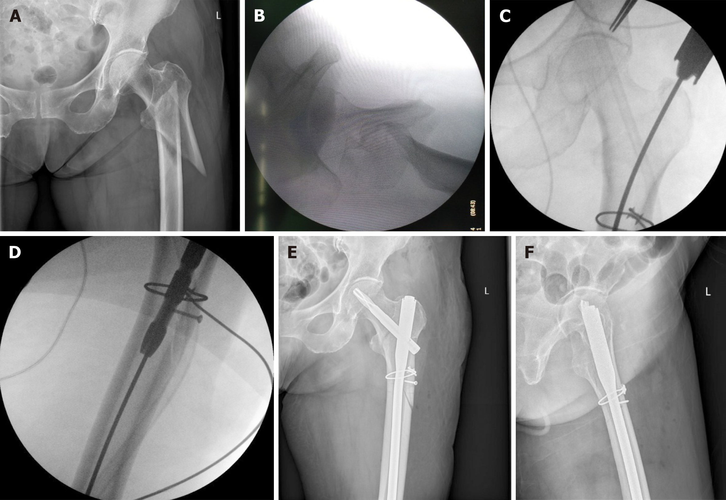
Figure 1 Typical presentation of a female patient, who was 78-years-old.
A: A radiograph of type 31A3 intertrochanteric fracture upon preoperative anteroposterior X-ray; B: A radiograph from intraoperative C-arm, which confirmed the irreducible intertrochanteric fracture; C-D: the insertion of proximal femoral nail antirotation without the need for Kirschner wires and other reduction instruments after limited open reduction with intracortical screws; E-F: The anatomical reduction of fracture upon anteroposterior and lateral X-rays.
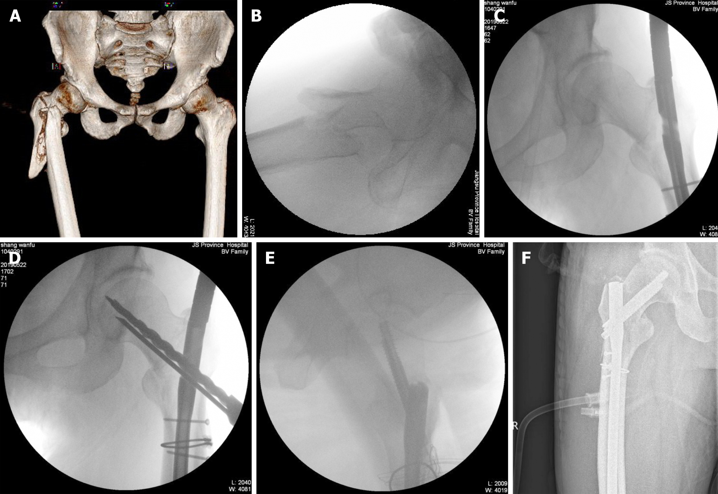
Figure 2 Typical presentation of a male patient, who was 74-years-old.
A: A preoperative 3D computed tomography reconstruction of type 31A3 intertrochanteric fracture; B: A radiograph from intraoperative C-arm, which confirmed the irreducible intertrochanteric fracture; C-E: The insertion of META-TAN without the need for Kirschner wires and other reduction instruments after limited open reduction with intracortical screws; F: The anatomical reduction of fracture upon postoperative anteroposterior X-ray.
- Citation: Huang XW, Hong GQ, Zuo Q, Chen Q. Intracortical screw insertion plus limited open reduction in treating type 31A3 irreducible intertrochanteric fractures in the elderly. World J Clin Cases 2021; 9(32): 9752-9761
- URL: https://www.wjgnet.com/2307-8960/full/v9/i32/9752.htm
- DOI: https://dx.doi.org/10.12998/wjcc.v9.i32.9752


