Copyright
©The Author(s) 2021.
World J Clin Cases. Nov 16, 2021; 9(32): 10040-10045
Published online Nov 16, 2021. doi: 10.12998/wjcc.v9.i32.10040
Published online Nov 16, 2021. doi: 10.12998/wjcc.v9.i32.10040
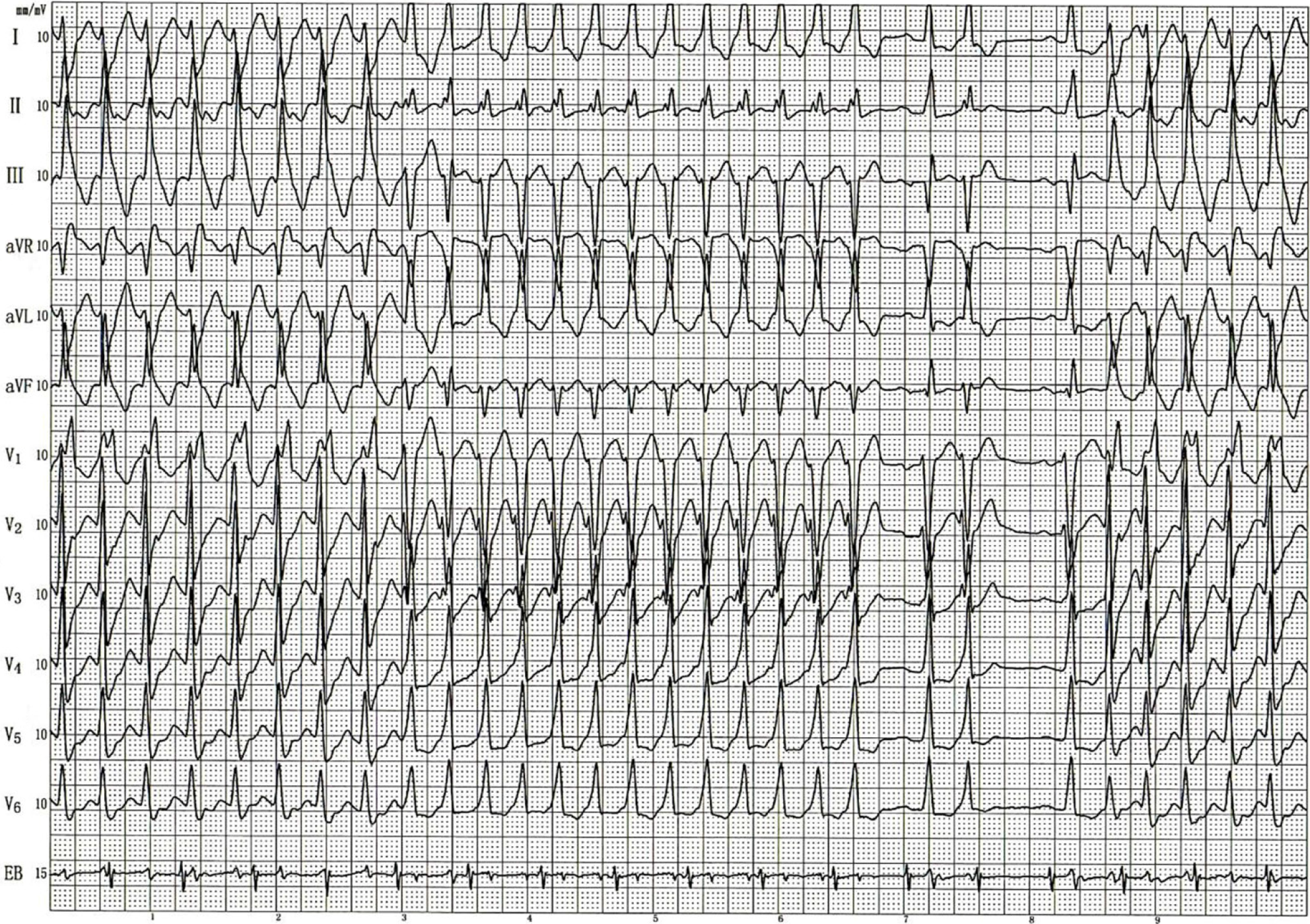
Figure 1 12-lead electrocardiogram obtained during a transesophageal electrophysiological study.
For the first several beats, the electrocardiogram pattern corresponded to a right-bundle-branch block and left-posterior-branch block, with a rate of 187 bpm. The pattern subsequently spontaneously changed to another form with a rate of 202 bpm, showing rS in lead V1, positive QRS polarity in leads I, II and aVL and negative QRS polarity in leads II, III and aVF. A few seconds later, sinus rhythm appeared and captured the His bundle potential. The next atrial premature contraction with a long interval induced a subsequent episode of ventricular tachycardia, similar to the first several beats. The ventricular potential was always matched with the His potential but was not related to the atrial potential; that is, the AA and VV intervals were constant.
- Citation: Zhang LY, Dong SJ, Yu HJ, Chu YJ. Ventricular tachycardia originating from the His bundle: A case report. World J Clin Cases 2021; 9(32): 10040-10045
- URL: https://www.wjgnet.com/2307-8960/full/v9/i32/10040.htm
- DOI: https://dx.doi.org/10.12998/wjcc.v9.i32.10040


