Copyright
©The Author(s) 2021.
World J Clin Cases. Nov 16, 2021; 9(32): 10006-10012
Published online Nov 16, 2021. doi: 10.12998/wjcc.v9.i32.10006
Published online Nov 16, 2021. doi: 10.12998/wjcc.v9.i32.10006
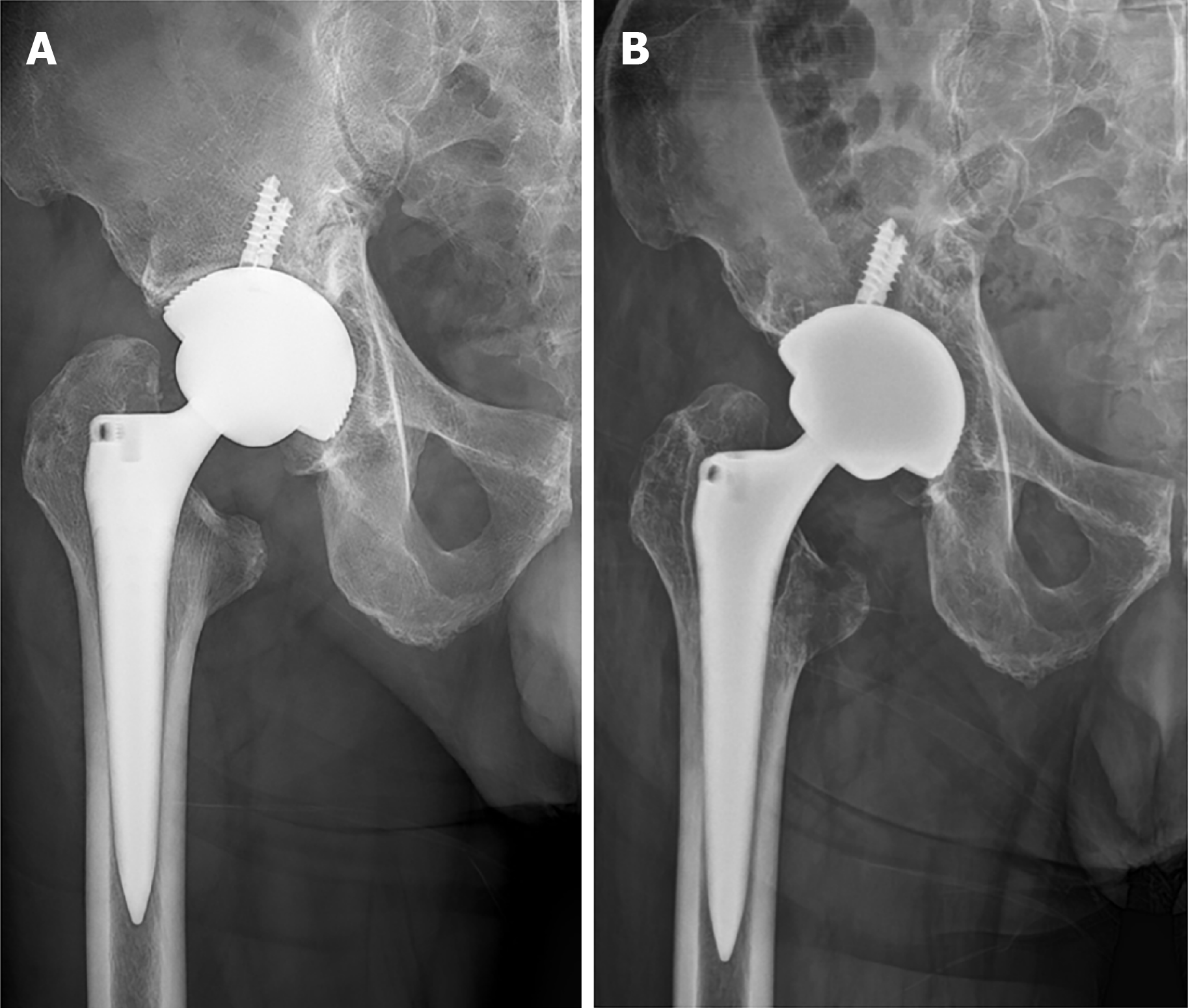
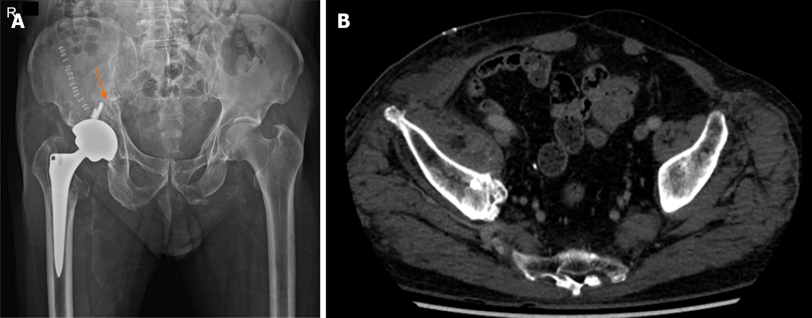
Figure 1 Anterolateral pelvis radiographs of the case.
A: Immediately following the index total hip arthroplasty; B: At the time of initial visit to our institution.
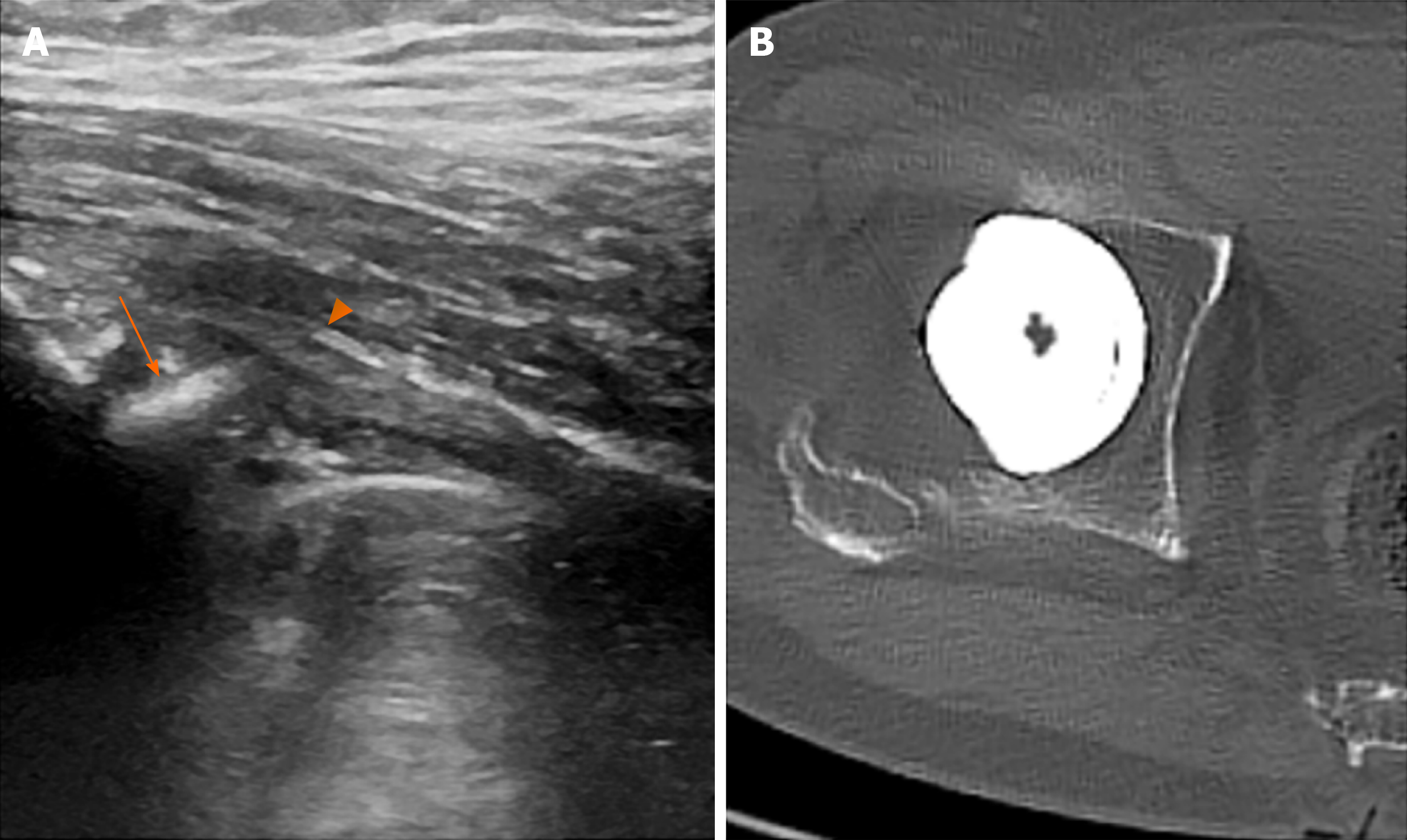
Figure 2 Images showing potential iliopsoas impingement by the cup.
A: Ultrasonography demonstrating a protruded acetabular rim (arrow) in approximation to the iliopsoas tendon (arrow head); B: Axial computed tomography scan showing a retroverted cup by 9°.
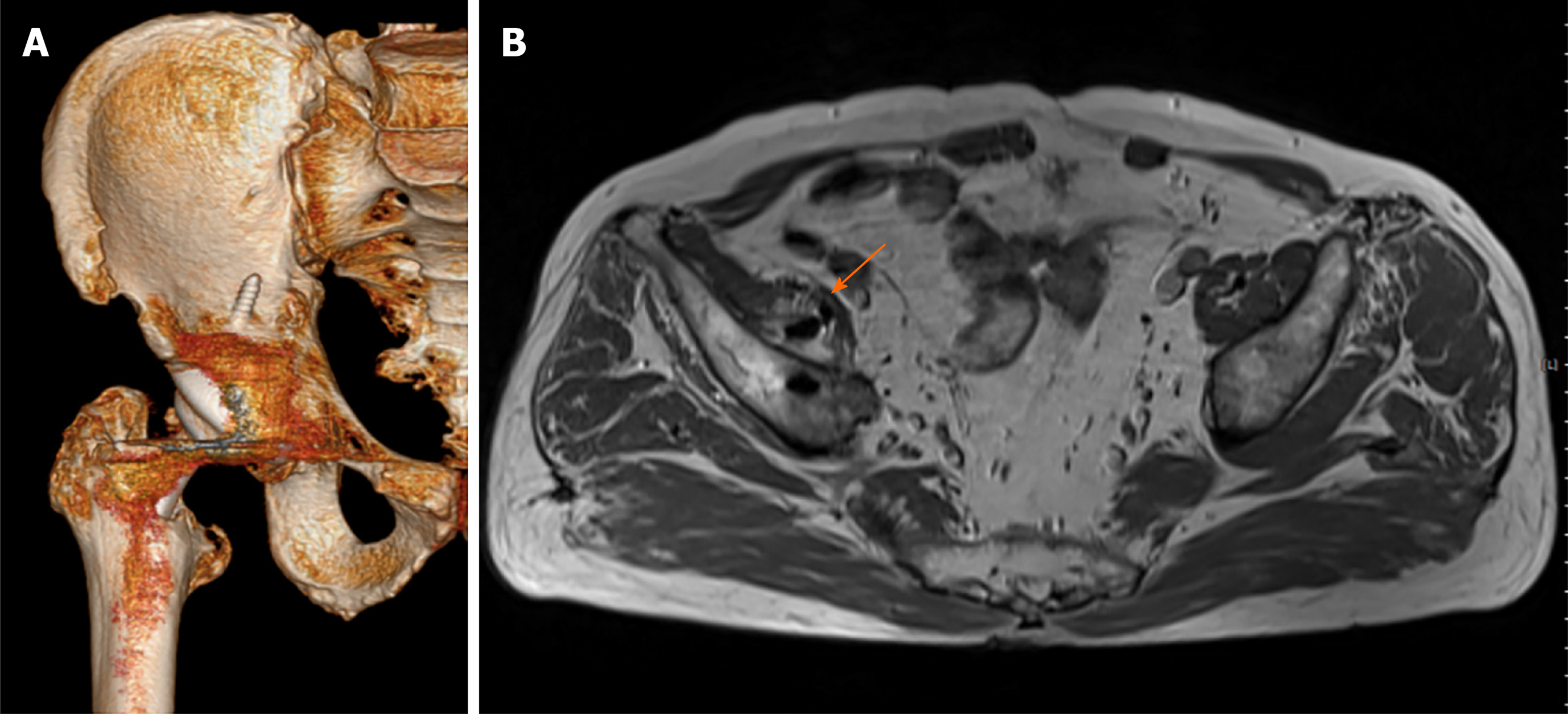
Figure 3 Advance images showing screw protrusion.
A: Three-dimensional reconstruction of a computed tomography image showing the screw penetrating through the ilium; B: Magnetic resonance imaging showing the screw penetrating through the iliopsoas muscle (arrow).
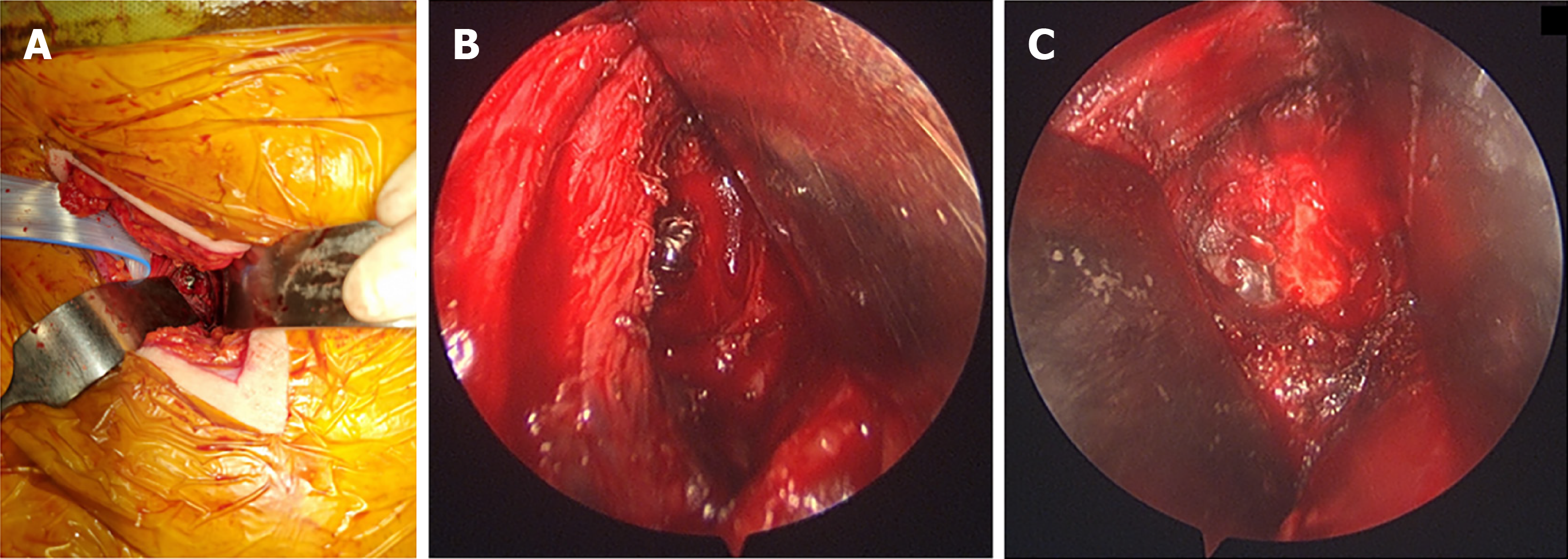
Figure 4 Clinical photos from surgical intervention.
A: Exposure of the screw using the pararectus approach; B: Close-up look of the protruding screw; C: Following flattening of the screw.
Figure 5 Immediate postoperative radiographs.
A: Showing the flattened screw tip (arrow); B: Complete resection of the screw (as compare to Figure 3B).
- Citation: Park HS, Lee SH, Cho HM, Choi HB, Jo S. Screw penetration of the iliopsoas muscle causing late-onset pain after total hip arthroplasty: A case report. World J Clin Cases 2021; 9(32): 10006-10012
- URL: https://www.wjgnet.com/2307-8960/full/v9/i32/10006.htm
- DOI: https://dx.doi.org/10.12998/wjcc.v9.i32.10006


