Copyright
©The Author(s) 2021.
World J Clin Cases. Nov 6, 2021; 9(31): 9680-9685
Published online Nov 6, 2021. doi: 10.12998/wjcc.v9.i31.9680
Published online Nov 6, 2021. doi: 10.12998/wjcc.v9.i31.9680
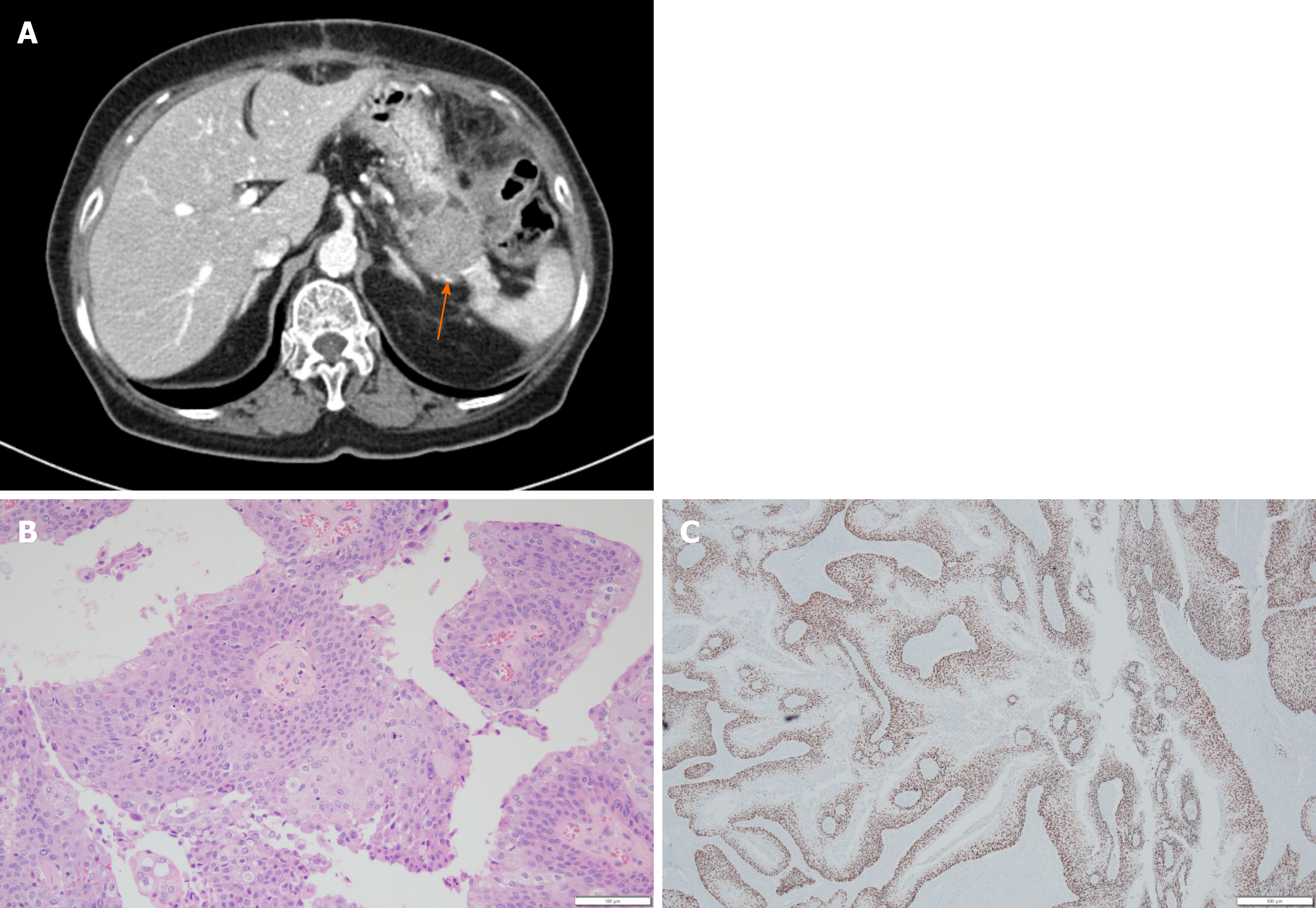
Figure 1 Images of the pancreas.
A: Computed tomography showing 3.3 cm sized mass (arrow) at the pancreas tail with enhancement; B: Hematoxylin & eosin stain of distal pancreas showing moderately differentiated squamous cell carcinoma with papillary growth pattern (× 200); C: Tumor cells were diffuse positive for p63 by immunohistochemical staining (× 40).
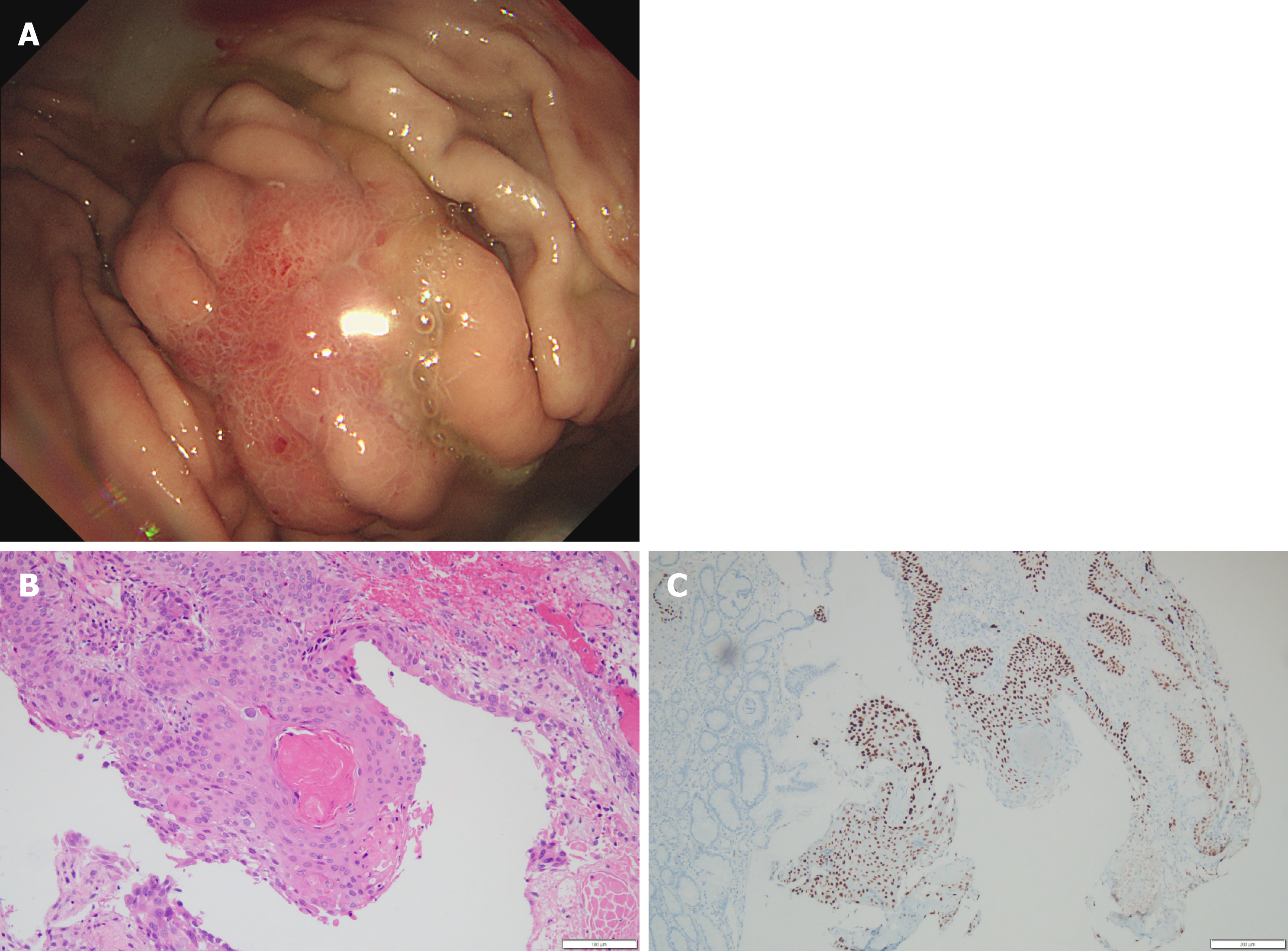
Figure 2 Images of the stomach.
A: Gastroscopy showing 2 cm sized ill-defined irregular flat and hyperemic mucosa at high body; B: Hematoxylin & eosin stain of stomach biopsy showing moderately differentiated squamous cell carcinoma (× 200); C: Tumor cells were positive for p63 by immunohistochemical staining (× 100).
- Citation: Kim JH, Kang CD, Lee K, Lim KH. Metachronous squamous cell carcinoma of pancreas and stomach in an elderly female patient: A case report. World J Clin Cases 2021; 9(31): 9680-9685
- URL: https://www.wjgnet.com/2307-8960/full/v9/i31/9680.htm
- DOI: https://dx.doi.org/10.12998/wjcc.v9.i31.9680


