Copyright
©The Author(s) 2021.
World J Clin Cases. Oct 26, 2021; 9(30): 9295-9301
Published online Oct 26, 2021. doi: 10.12998/wjcc.v9.i30.9295
Published online Oct 26, 2021. doi: 10.12998/wjcc.v9.i30.9295
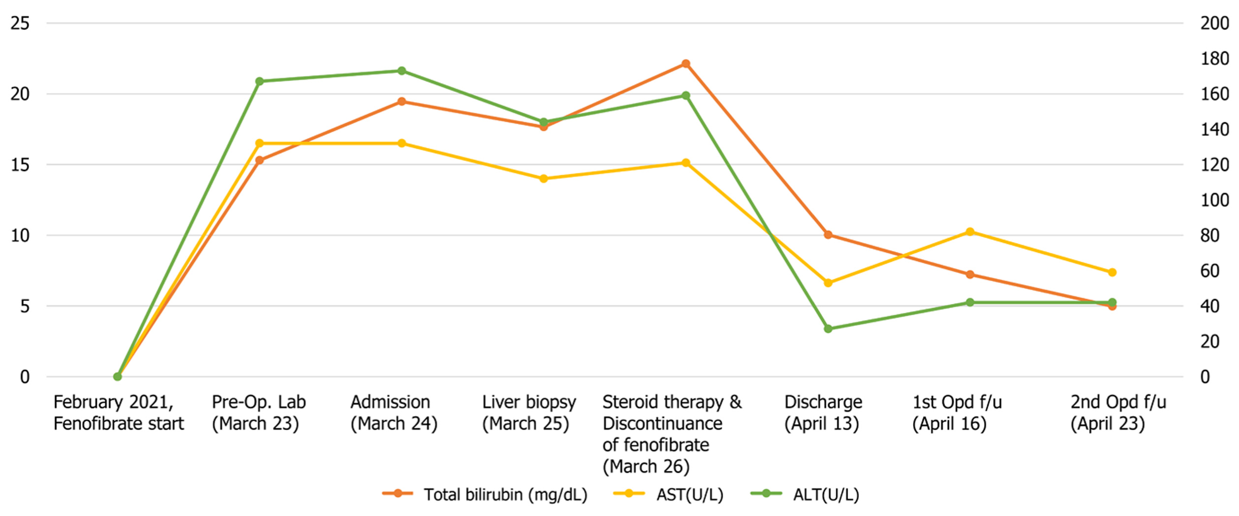
Figure 1 Clinical course of the patient.
AST: Aspartate aminotransferase; ALT: Alanine aminotransferase.
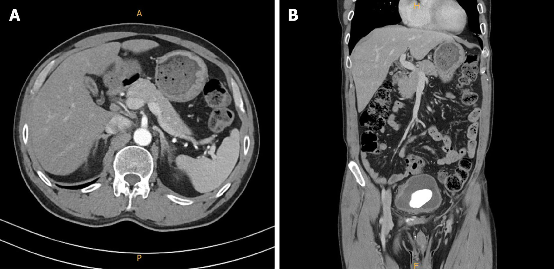
Figure 2 Computed tomography of the patient.
Contrast-enhanced abdominal computed tomography revealed no bile duct obstruction. A: Transverse plane; B: Coronal view.
Figure 3 Results of liver biopsy.
Magnification: 400 ×; scale bar: 25 μm. A: The overall findings of sinusoidal inflammation in the portal tract increased. Hepatocytes are pinkish and ballooned. Portal vein fibrosis and increased nodular activity are found. Bile pigment is deposited in cytoplasm, resulting in a yellow tinge; B: Central vein is visible and cholestasis necrosis is concentrated around it. The lobule is concentrated in zone 3 with typical findings of acute cholestatic hepatitis.
- Citation: Lee HY, Lee AR, Yoo JJ, Chin S, Kim SG, Kim YS. Biopsy-confirmed fenofibrate-induced severe jaundice: A case report. World J Clin Cases 2021; 9(30): 9295-9301
- URL: https://www.wjgnet.com/2307-8960/full/v9/i30/9295.htm
- DOI: https://dx.doi.org/10.12998/wjcc.v9.i30.9295


