Copyright
©The Author(s) 2021.
World J Clin Cases. Oct 26, 2021; 9(30): 9285-9294
Published online Oct 26, 2021. doi: 10.12998/wjcc.v9.i30.9285
Published online Oct 26, 2021. doi: 10.12998/wjcc.v9.i30.9285
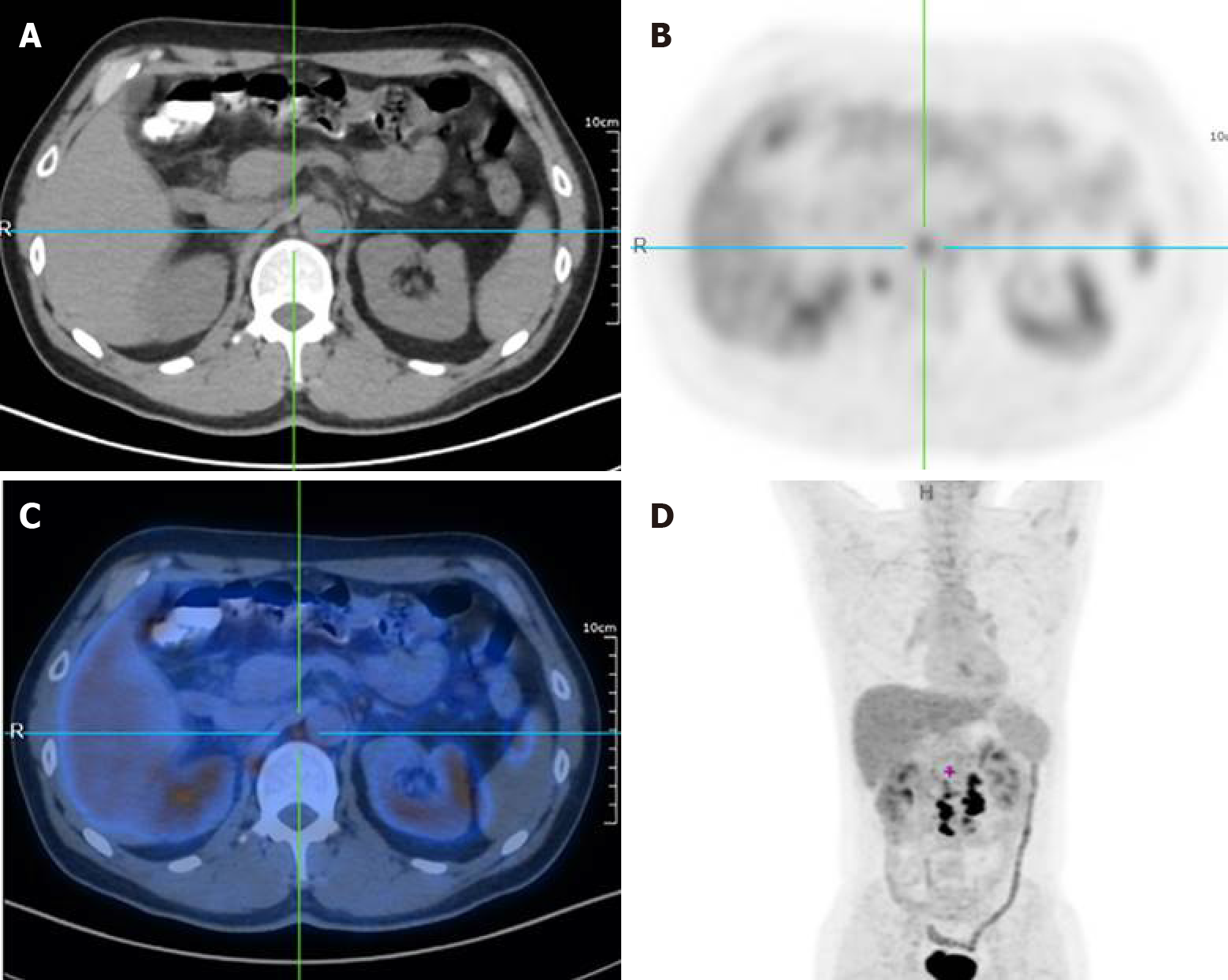
Figure 1 Multiple lymph nodes metastases around the abdominal aorta.
A: Computed tomography (CT) image. The red arrow points to an enlarged lymph node adjacent to the abdominal aorta; B: 18F-fluorodeoxyglucose (18F-FDG) positron emission tomography (PET)-CT image. The center of the picture is an enlarged lymph node adjacent to the abdominal aorta, and the standard uptake value in this part is significantly increased; C: 18F-FDG PET-CT image. Figure 2B and Figure 2C are overlapped; D: Whole body 18F-FDG PET-CT image. The center of the red cross is an enlarged lymph node adjacent to the abdominal aorta.
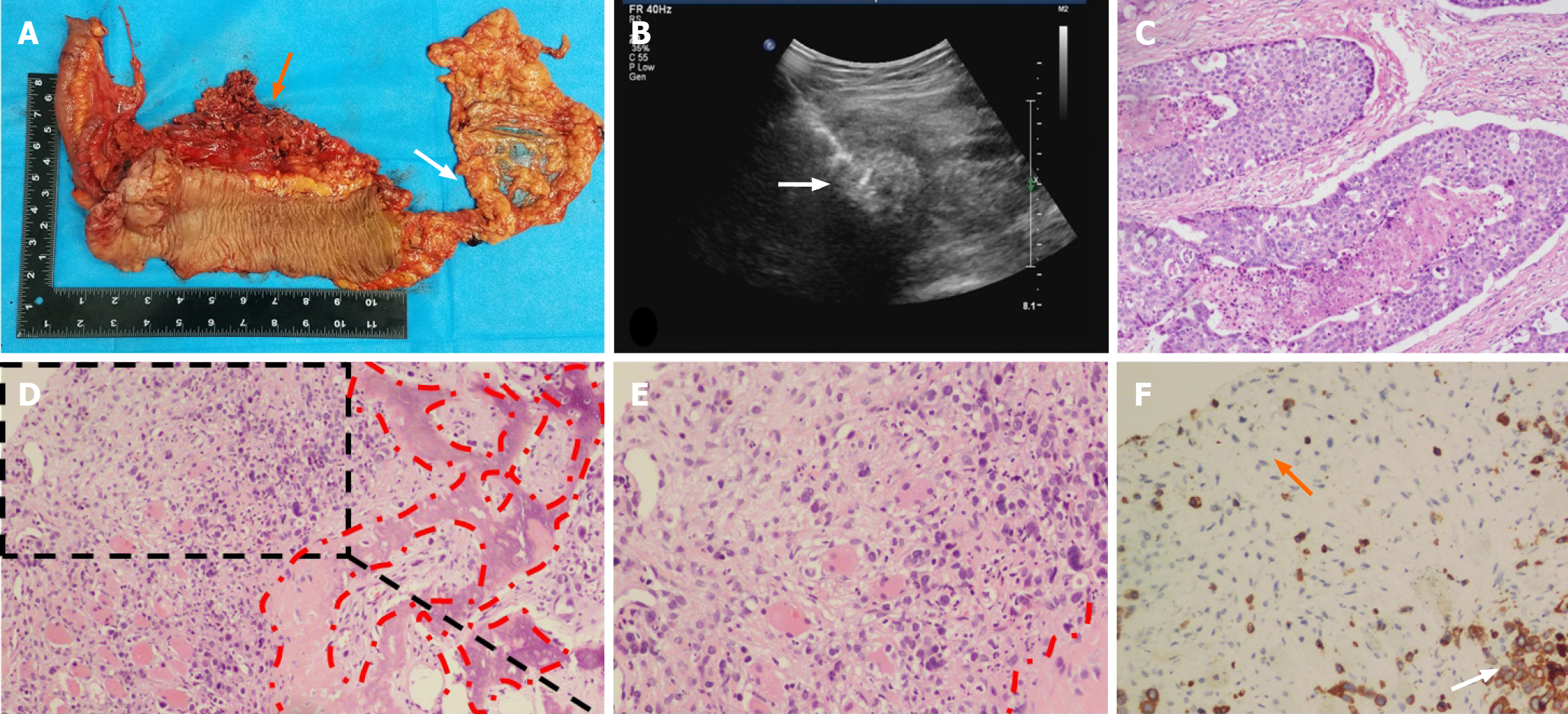
Figure 2 Pathology and immunohistochemistry of the primary lesion and thigh mass.
A: In the resected specimen, the orange arrow points to the tumor location and the white arrow points to the appendix; B: Skeletal muscle metastasis (white arrow) on color ultrasound; C: Hematoxylin-eosin staining of the resected colon cancer specimen (magnification, 40 times); D: H&E staining of an SMM biopsy. The red dashed circle is the bone metaplasia region (magnification, 40 times); E: Part of the enlarged Figure 1D (magnification, 200 times); F: Cytokeratin staining of an SMM biopsy. The orange arrow refers to the fibroblasts in the tumor stroma and the white arrow refers to the metastatic tumor cells (magnification, 200 times).
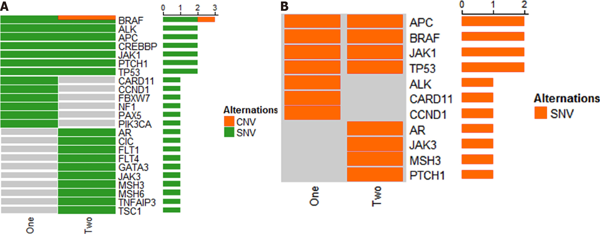
Figure 3 BRAF gene mutation detected using gene test.
A: The difference in gene expression of sequencing results; B: BRAF gene mutation detected using gene test. CNV: Copy number variation; SNV: Single nucleotide variation.
- Citation: Guo Y, Wang S, Zhao ZY, Li JN, Shang A, Li DL, Wang M. Skeletal muscle metastasis with bone metaplasia from colon cancer: A case report and review of the literature. World J Clin Cases 2021; 9(30): 9285-9294
- URL: https://www.wjgnet.com/2307-8960/full/v9/i30/9285.htm
- DOI: https://dx.doi.org/10.12998/wjcc.v9.i30.9285


