Copyright
©The Author(s) 2021.
World J Clin Cases. Oct 26, 2021; 9(30): 9218-9227
Published online Oct 26, 2021. doi: 10.12998/wjcc.v9.i30.9218
Published online Oct 26, 2021. doi: 10.12998/wjcc.v9.i30.9218
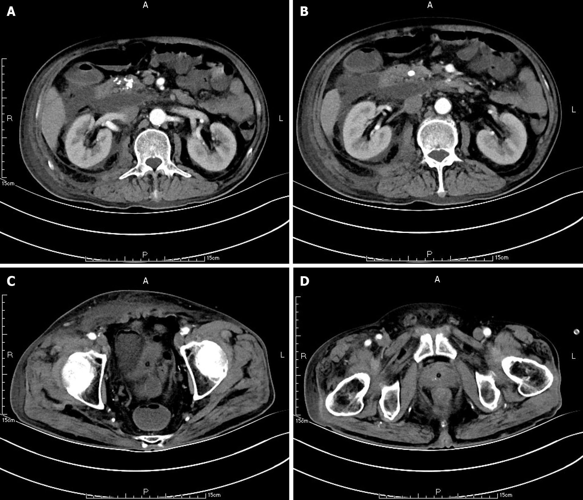
Figure 1 The outline of the uncinate process of the head of pancreas is unclear (A, B), and a large amount of exudation can be seen around the pancreas (C, D).
Pancreatic parenchyma atrophy, multiple calcification spots and pancreatic duct widening were observed. There was a small amount of subcutaneous and intramuscular effusion in the right abdominal wall.
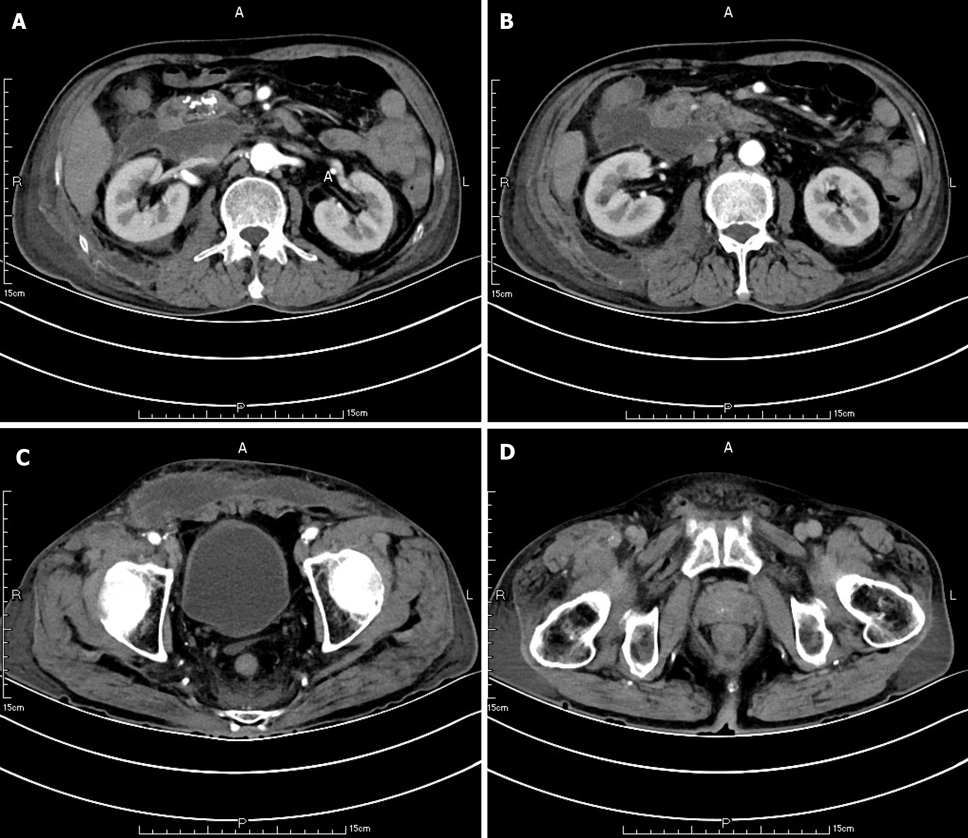
Figure 2 The range of encapsulated liquid density shadow in the right anterior (A, B), and posterior renal space was slightly larger than that in the front and extended to the right iliac fossa (C, D).
The subcutaneous and intramuscular effusion of abdominal wall increased.
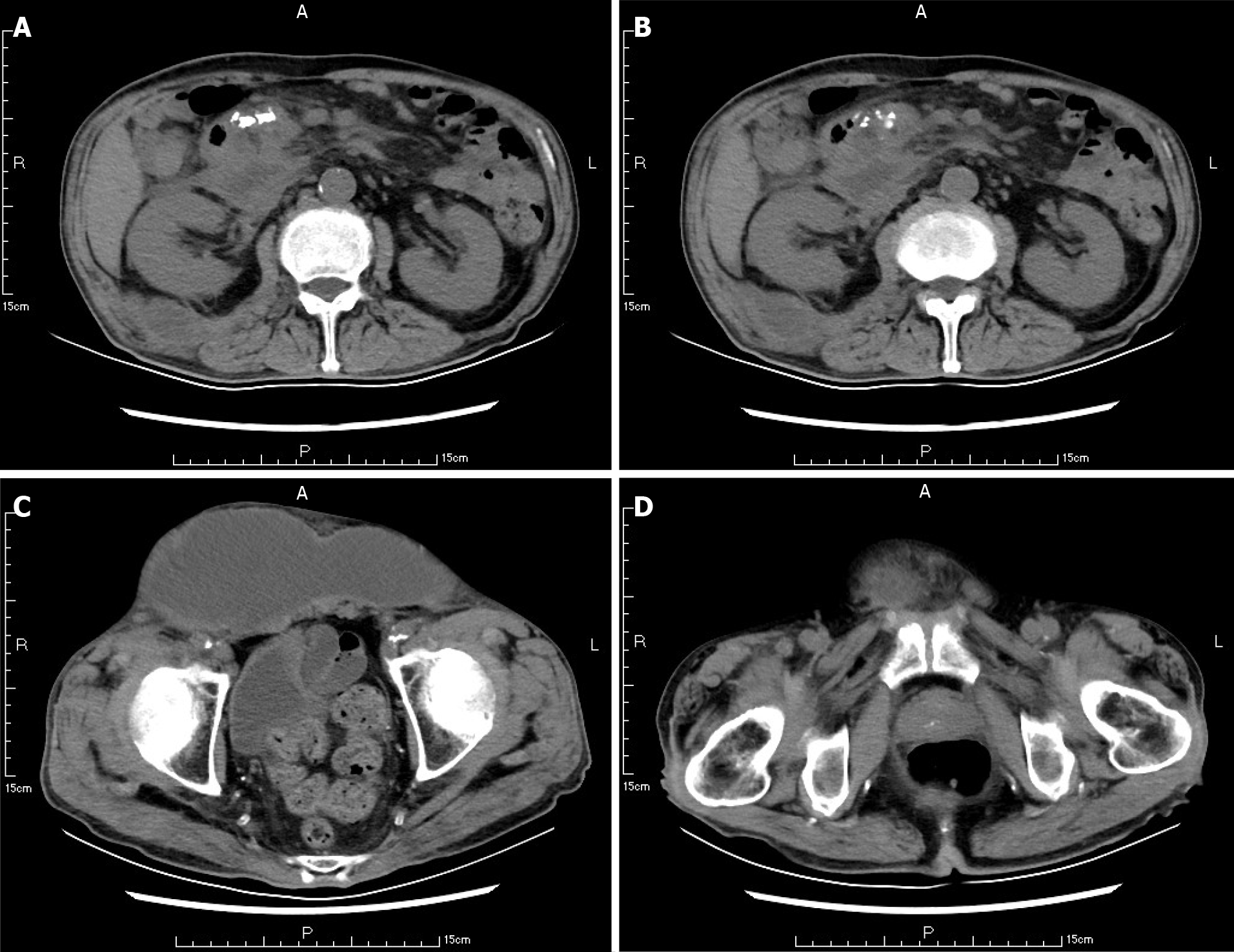
Figure 3 The range of encapsulated liquid density shadow in the right retrorenal space was significantly larger than that before (A, B), extending to the right iliac fossa (C, D).
The density range of subcutaneous and intramuscular fluid in the right abdominal wall was significantly larger than that before. Right inguinal fluid density shadow.
Figure 4
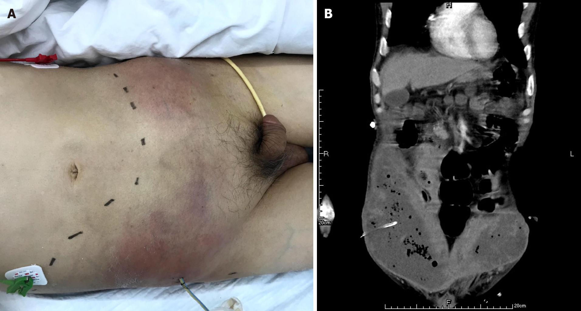
One abdominal wall drainage tube was placed after the first puncture and drainage (A, B).
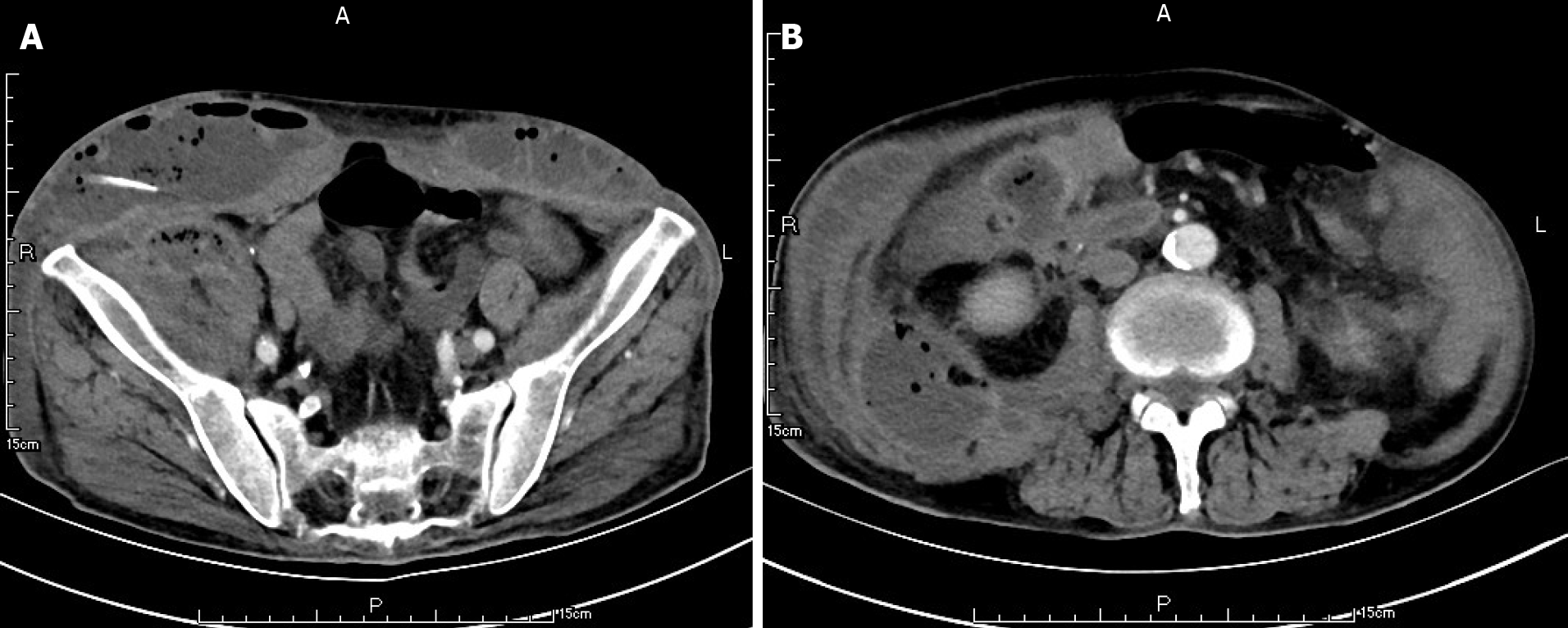
Figure 5 After the first puncture and drainage, there was encapsulated hydrops and pneumatosis in the abdominal wall (A, B).
There was extensive encapsulated hydrops and pneumatosis in the right retrorenal space.
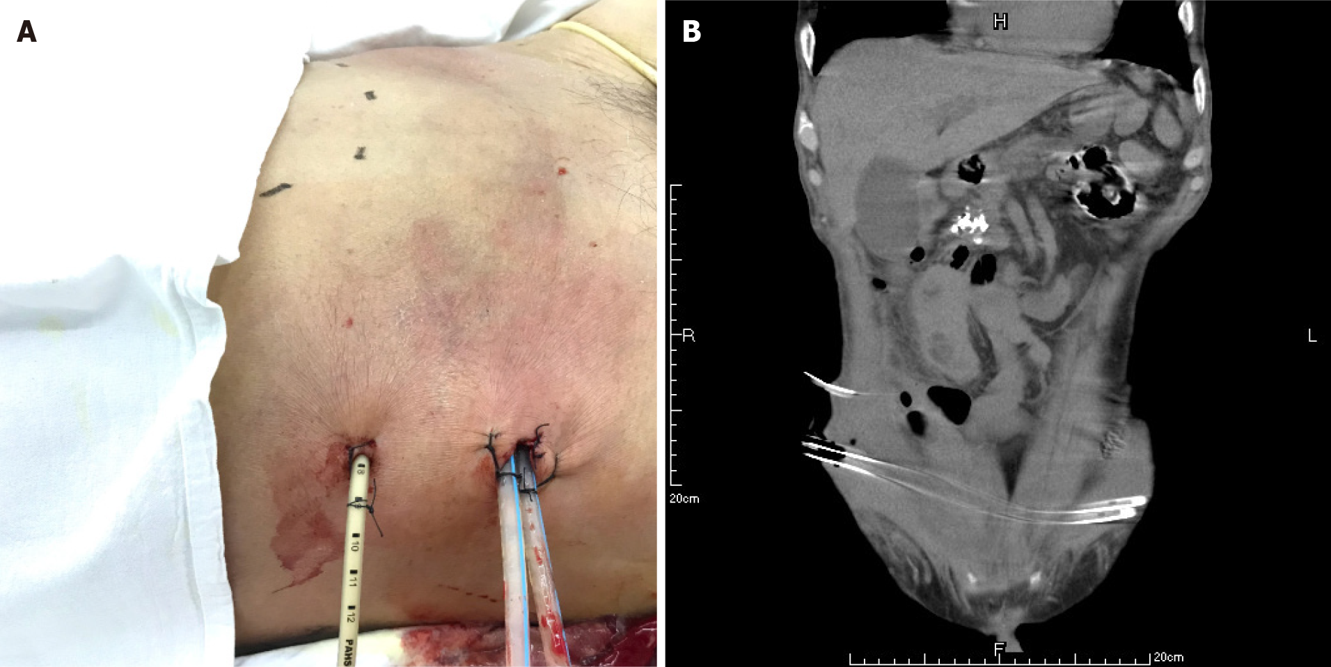
Figure 6
One retroperitoneal drainage tube and two abdominal wall drainage tubes were placed after the second puncture and drainage (A, B).
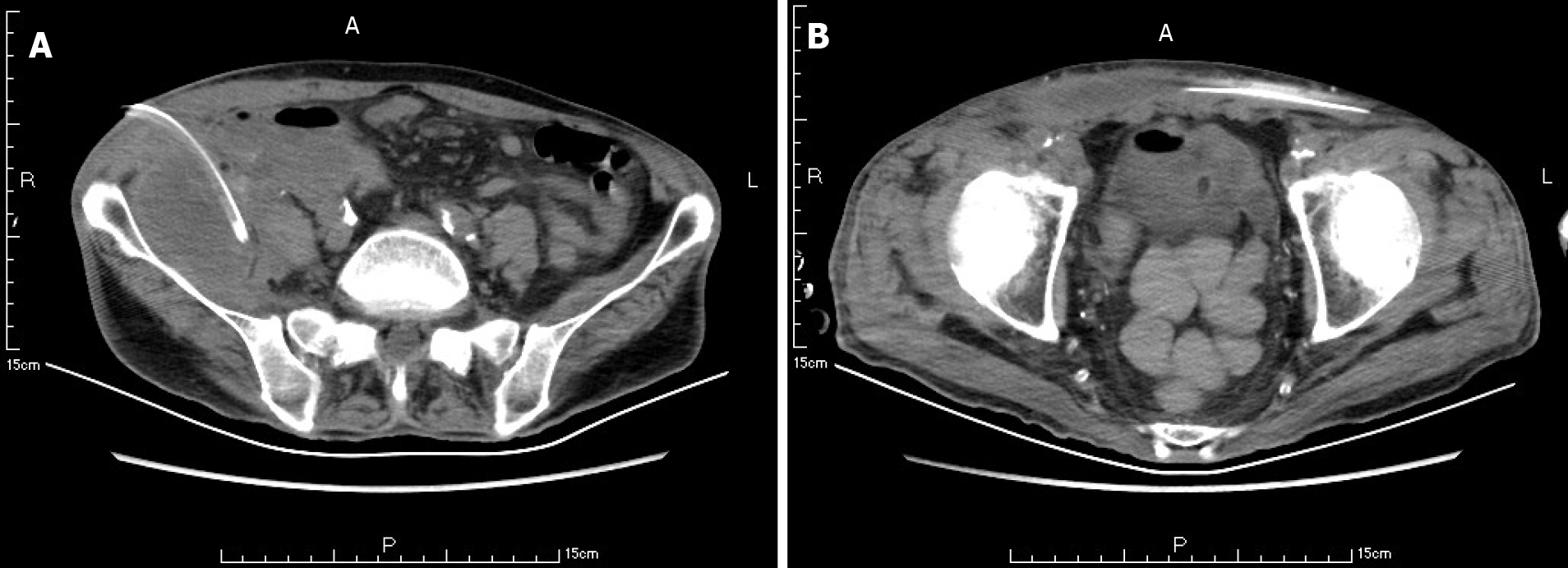
Figure 7 The subcutaneous and intramuscular effusion and pneumatosis of anterior abdominal wall were significantly reduced (A, B).
The arrow shows the retroperitoneal drainage tube, and the arrow shows the abdominal wall drainage tube.
- Citation: Jia YC, Ding YX, Mei WT, Xue ZG, Zheng Z, Qu YX, Li J, Cao F, Li F. Anterior abdominal abscess - a rare manifestation of severe acute pancreatitis: A case report. World J Clin Cases 2021; 9(30): 9218-9227
- URL: https://www.wjgnet.com/2307-8960/full/v9/i30/9218.htm
- DOI: https://dx.doi.org/10.12998/wjcc.v9.i30.9218


