Copyright
©The Author(s) 2021.
World J Clin Cases. Oct 26, 2021; 9(30): 9134-9143
Published online Oct 26, 2021. doi: 10.12998/wjcc.v9.i30.9134
Published online Oct 26, 2021. doi: 10.12998/wjcc.v9.i30.9134
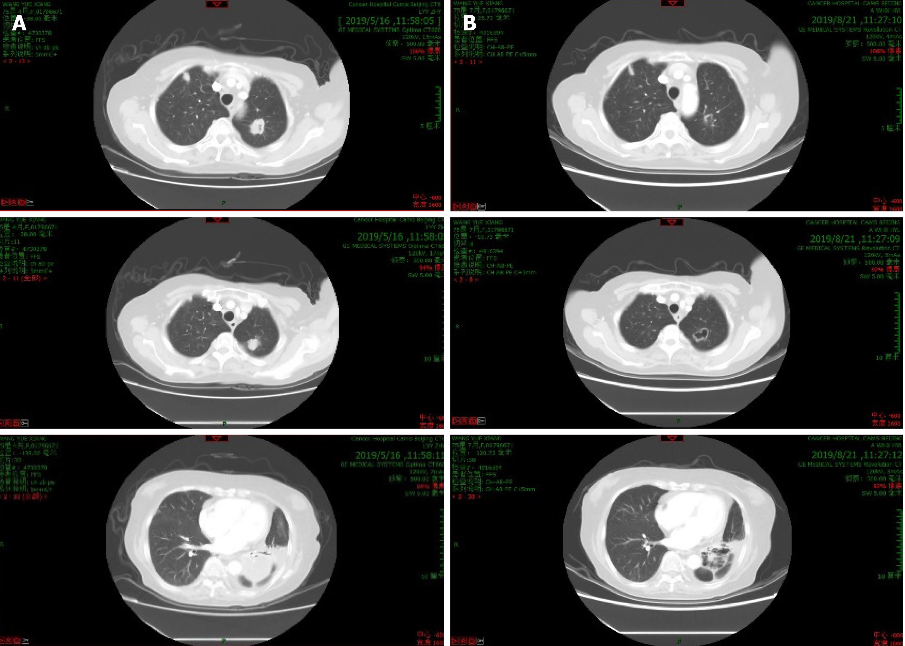
Figure 1 Changes in lung lesions after nab-paclitaxel plus apatinib treatment.
A: Computed tomography (CT) before treatment (baseline); B: CT scans after 6 cycles of nab-paclitaxel plus apatinib.
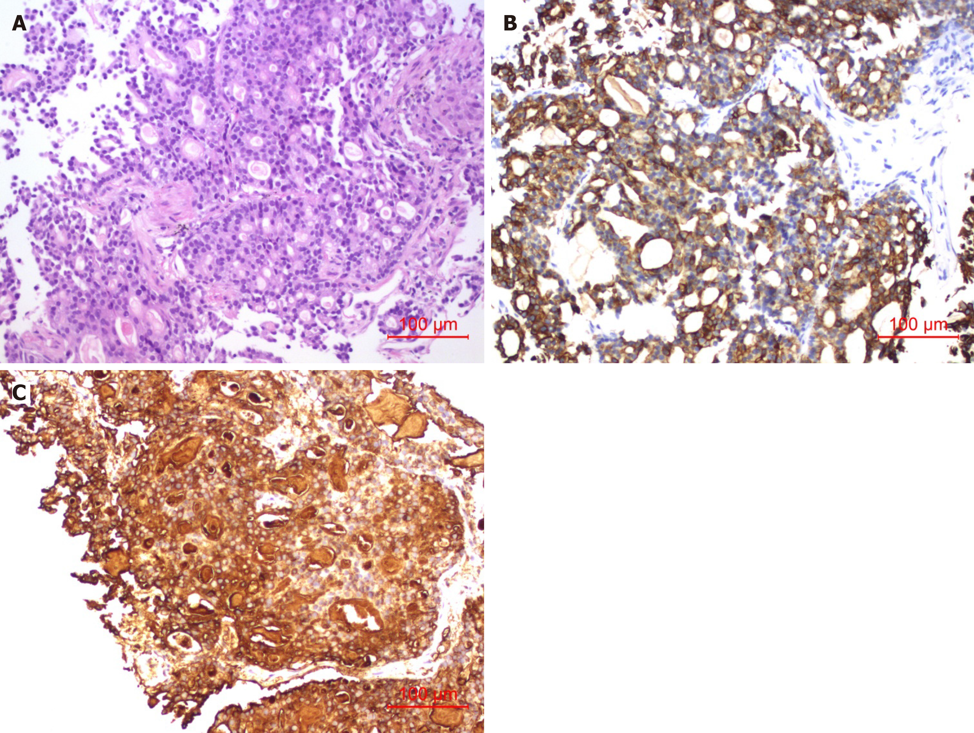
Figure 2 Needle biopsy of lung metastasis.
A: Hematoxylin and eosin stain of tissue with tubular structures, and cells that are not significantly heteromorphic (original magnification × 200); B: Immunohistochemical staining of CK19 was strongly positive on tumor-cell membranes (original magnification × 200); C: Immunohistochemical staining of CA19-9 was strongly positive in tumor-cell cytoplasm (original magnification × 200).
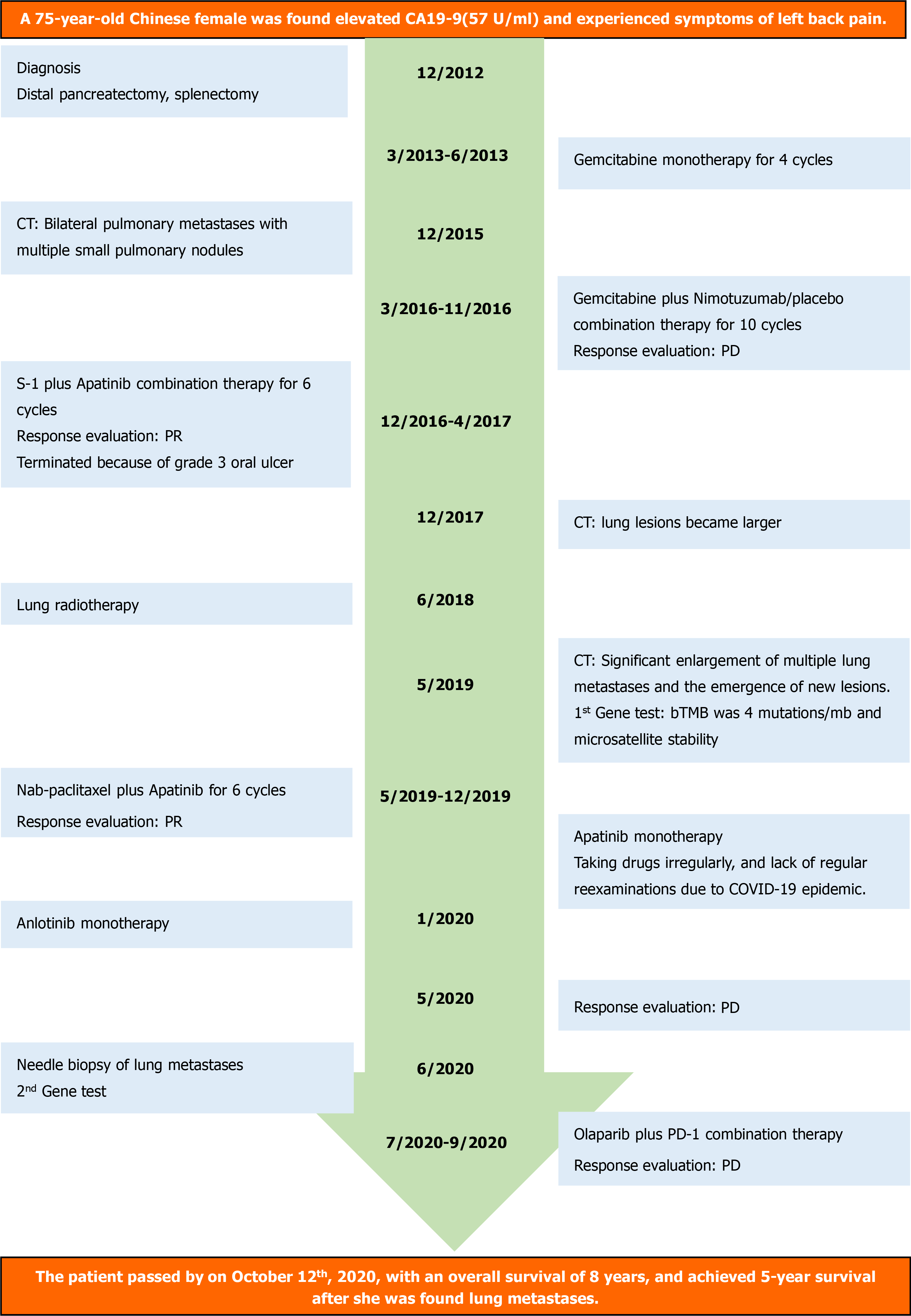
Figure 3 Timeline of interventions and outcomes.
bTMB: Blood tumor mutational burden; CA19-9: Carbohydrate antigen 19-9; CT: Computed tomography; COVID-19: Coronavirus disease 2019; PD: Progressive disease; PR: Partial response.
- Citation: Yang WW, Yang L, Lu HZ, Sun YK. Long-term survival of a patient with pancreatic cancer and lung metastasis: A case report and review of literature. World J Clin Cases 2021; 9(30): 9134-9143
- URL: https://www.wjgnet.com/2307-8960/full/v9/i30/9134.htm
- DOI: https://dx.doi.org/10.12998/wjcc.v9.i30.9134


