Copyright
©The Author(s) 2021.
World J Clin Cases. Oct 26, 2021; 9(30): 9114-9121
Published online Oct 26, 2021. doi: 10.12998/wjcc.v9.i30.9114
Published online Oct 26, 2021. doi: 10.12998/wjcc.v9.i30.9114
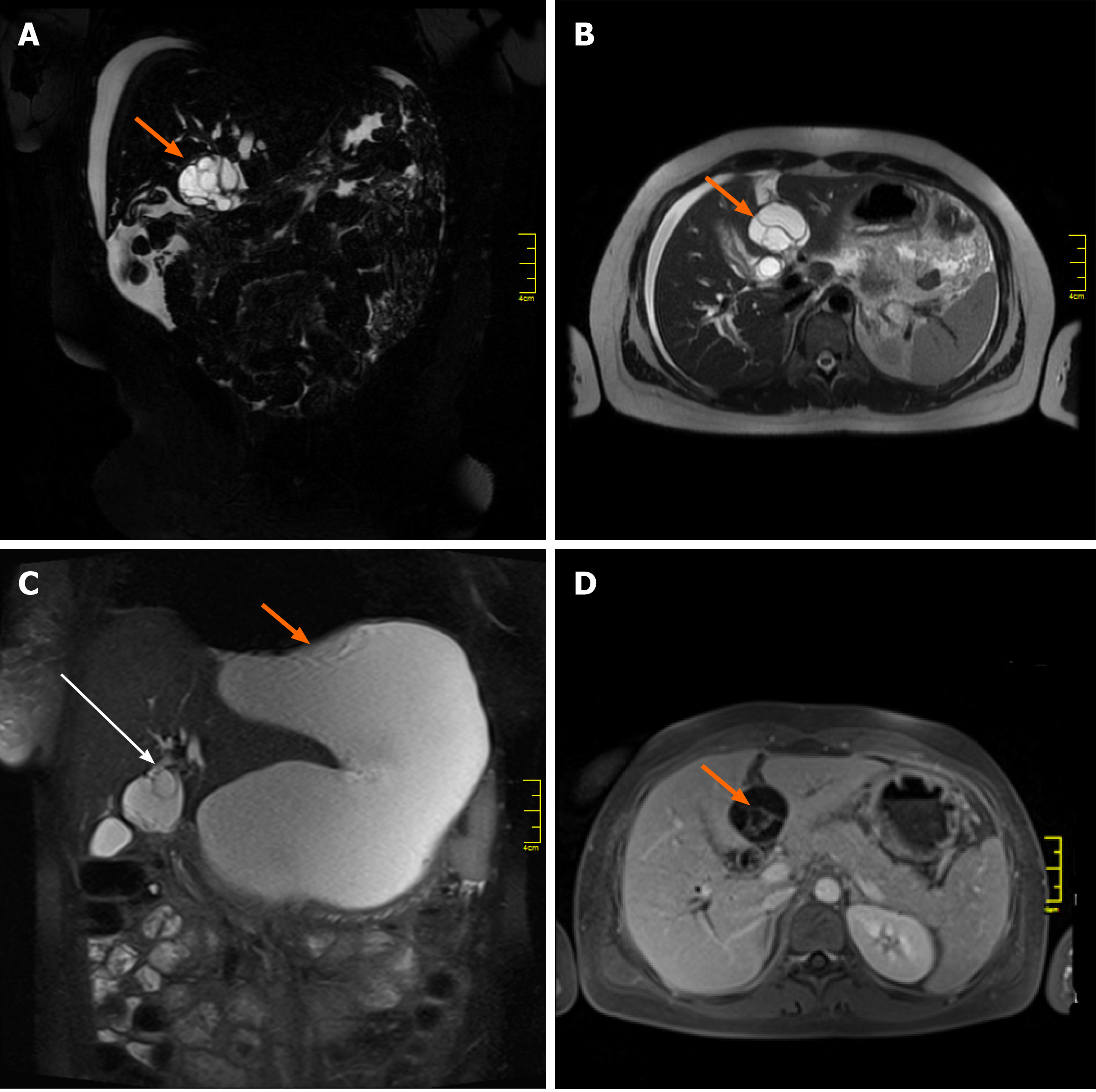
Figure 1 Magnetic resonance imaging studies of the cystic mass in the liver.
A, B: Initial magnetic resonance imaging (MRI) coronal and axial T2-weighted images showing a multilocular cystic mass (arrow), located between the left lateral segments of the liver (2/3) and segment 4; C: MRI T2-weighted coronal image demonstrating communication of the cystic mass with the left hepatic duct (long arrow) and a large fluid collection (short arrow) located anteriorly; D: MRI axial T1 image after intravenous gadolinium contrast administration, showing enhancement of internal septations (arrow) within the cystic liver mass.
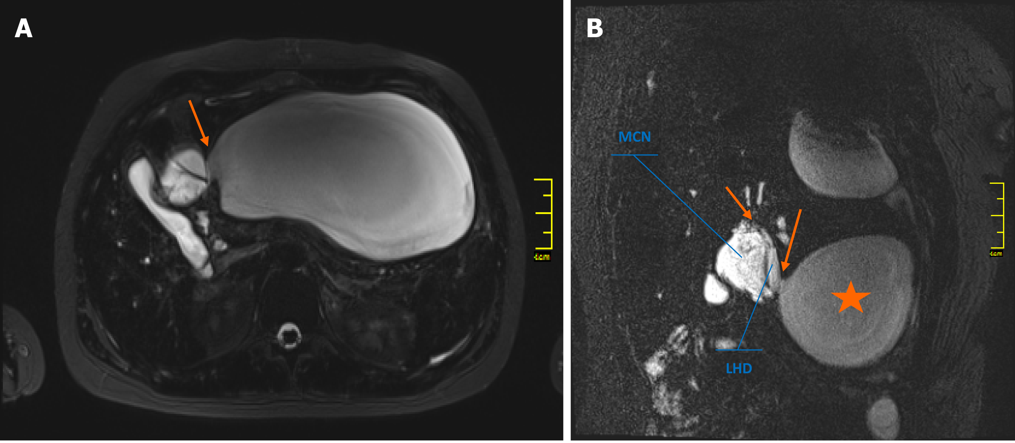
Figure 2 Communication of the mucinous cystic neoplasm and local structures.
A: Magnetic resonance imaging axial T2-weighted image showing the fluid collection directly adjacent to the mucinous cystic neoplasm (MCN) (arrow); B: The communication (arrows) between the MCN, left hepatic duct, and fluid collection (star) was better depicted on oblique coronal T2-weighted images. MCN: Mucinous cystic neoplasm; LHD: Left hepatic duct.
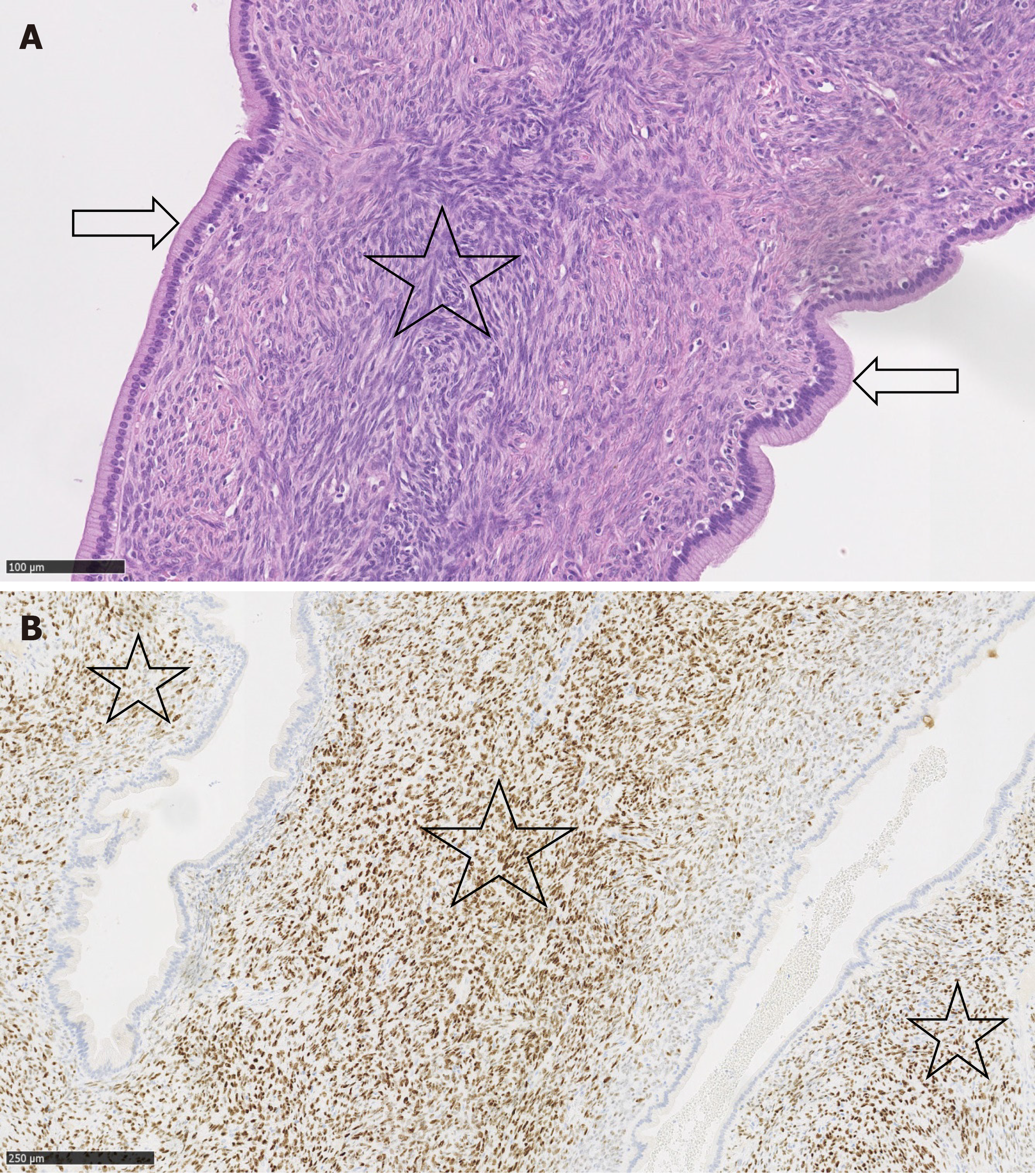
Figure 3 Mucinous cystic neoplasm.
A: Cystic spaces lined by single-layered columnar mucus-secreting epithelium without cytological atypia (arrow). The epithelium is surrounded by dense ovarian-type stroma (Star) (20×, haematoxylin and eosin stain); B: Progesterone receptors are expressed in dense, ovarian-type stroma (Star) (10× progesterone).
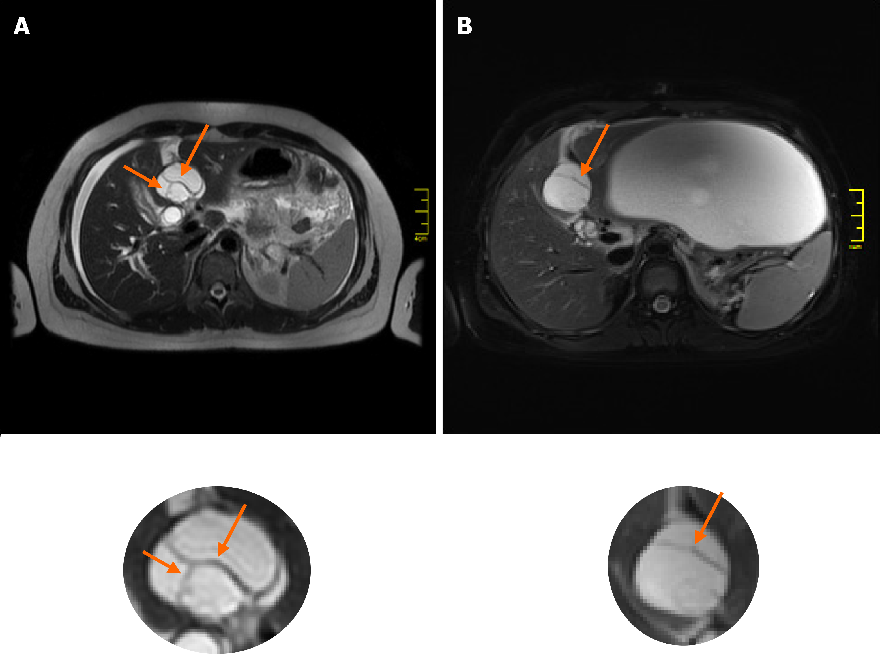
Figure 4 Changes in internal features of the cyst between magnetic resonance imaging studies.
Comparing the baseline magnetic resonance imaging (MRI) study (A) with the post-rupture MRI examination (B), the shape of the mucinous cystic neoplasm changed from oval to more circular. The internal architecture of its internal septations changed as demonstrated below (arrows).
- Citation: Kośnik A, Stadnik A, Szczepankiewicz B, Patkowski W, Wójcicki M. Spontaneous rupture of a mucinous cystic neoplasm of the liver resulting in a huge biloma in a pregnant woman: A case report. World J Clin Cases 2021; 9(30): 9114-9121
- URL: https://www.wjgnet.com/2307-8960/full/v9/i30/9114.htm
- DOI: https://dx.doi.org/10.12998/wjcc.v9.i30.9114


