Copyright
©The Author(s) 2021.
World J Clin Cases. Oct 26, 2021; 9(30): 9108-9113
Published online Oct 26, 2021. doi: 10.12998/wjcc.v9.i30.9108
Published online Oct 26, 2021. doi: 10.12998/wjcc.v9.i30.9108
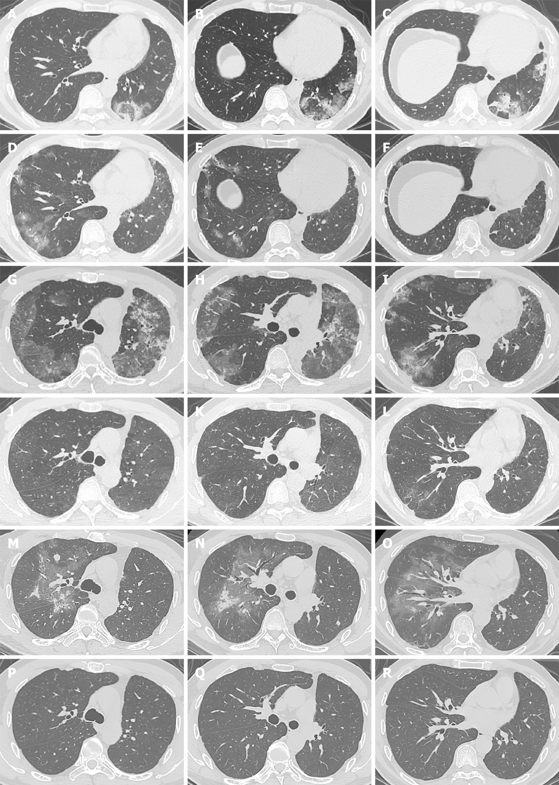
Figure 1 Computed tomography scan images of the patient during checkpoint inhibitor-related pneumonitis presentation and follow-up.
A-C: The first episode of checkpoint inhibitor-related pneumonitis (CIP) after 10 cycles of durvalumab; D-F: Chest computed tomography showed that the changes of the former CIP in the left upper lobe disappeared after 12 cycles of durvalumab; G-I: The second episode of CIP after 12 cycles of durvalumab; J-L: A significant improvement in the CIP at 1 wk after starting methylprednisolone; M-O: The third episode of CIP at completion of methylprednisolone tapering; P-R: Resolution of CIP at 5 mon after completion of methylprednisolone tapering.
Figure 2
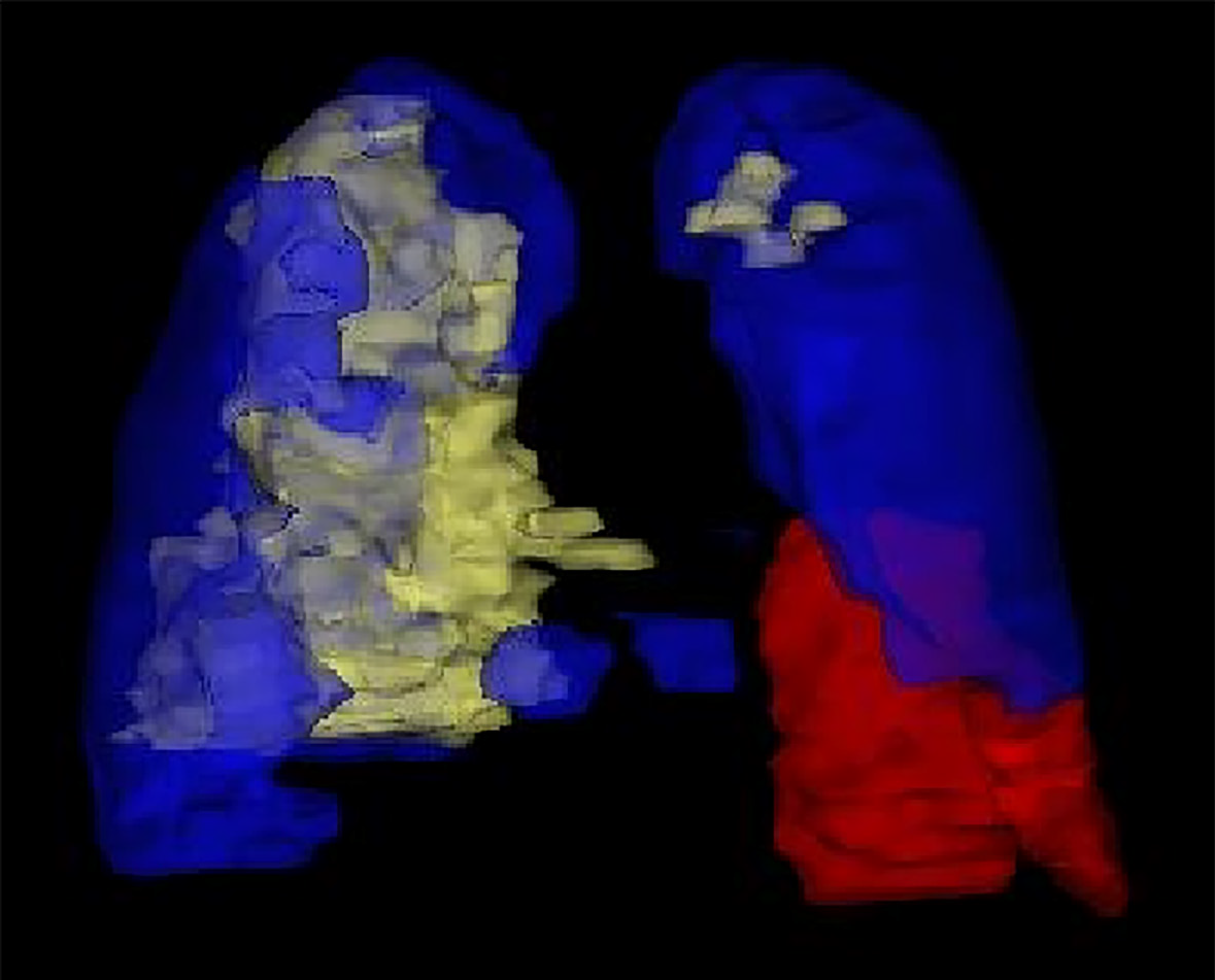
A three-dimensional reconstruction of the pneumonitis areas for three episodes.
The red, blue, and yellow areas show the first, second, and third episodes of checkpoint inhibitor-related pneumonitis, respectively, taking the pneumonitis areas all together makes nearly a whole lung.
- Citation: Tan PX, Huang W, Liu PP, Pan Y, Cui YH. Dynamic changes in the radiologic manifestation of a recurrent checkpoint inhibitor related pneumonitis in a non-small cell lung cancer patient: A case report. World J Clin Cases 2021; 9(30): 9108-9113
- URL: https://www.wjgnet.com/2307-8960/full/v9/i30/9108.htm
- DOI: https://dx.doi.org/10.12998/wjcc.v9.i30.9108


