Copyright
©The Author(s) 2021.
World J Clin Cases. Oct 16, 2021; 9(29): 8946-8952
Published online Oct 16, 2021. doi: 10.12998/wjcc.v9.i29.8946
Published online Oct 16, 2021. doi: 10.12998/wjcc.v9.i29.8946
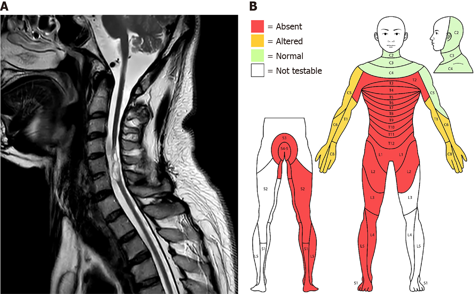
Figure 1 Cervical magnetic resonance imaging and sensory impairment point distribution of patients.
A: Severe volume loss of the spinal cord with cystic change (myelomalacia at the C5 and C6 vertebra level) was observed on magnetic resonance imaging at the time of admission with autonomic dysreflexia; B: Evaluation according to the International Standards for Neurological Classification of spinal cord injury. In the sensory test, anesthesia was observed in the region below T1. The patient underwent transfemoral amputation on the left lower extremity 16 years before.
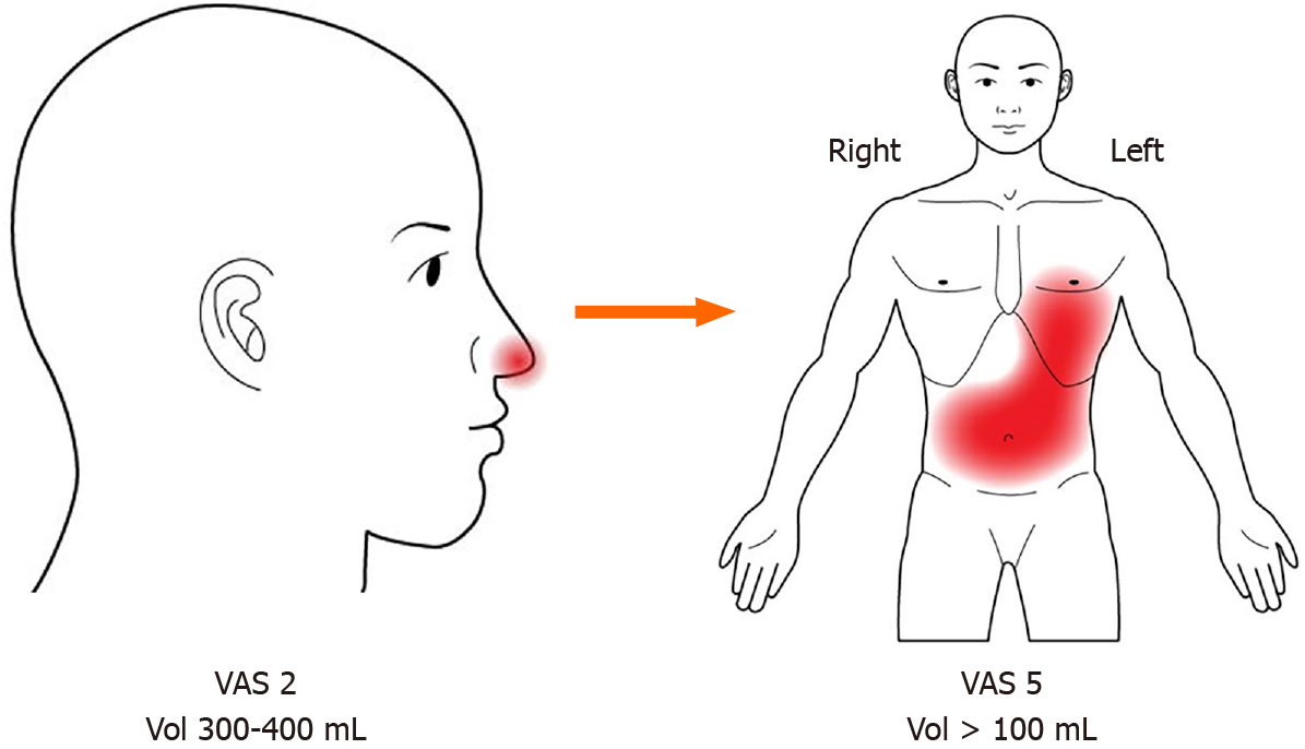
Figure 2 Changes in perception of bladder filling: Location, intensity, precision.
After the onset of autonomic dysreflexia, the location of the senses for bladder fullness (nose → abdominal, precordial area) and the degree of discomfort [visual analog scale (VAS) 2 → VAS 5] changed. In addition, the bladder volume for sense of fullness became smaller and variable (300–400 mL → > 100 mL). VAS: Visual analog scale.
- Citation: Yoon JY, Kim DS, Kim GW, Won YH, Park SH, Ko MH, Seo JH. Disruption of sensation-dependent bladder emptying due to bladder overdistension in a complete spinal cord injury: A case report. World J Clin Cases 2021; 9(29): 8946-8952
- URL: https://www.wjgnet.com/2307-8960/full/v9/i29/8946.htm
- DOI: https://dx.doi.org/10.12998/wjcc.v9.i29.8946


