Copyright
©The Author(s) 2021.
World J Clin Cases. Oct 16, 2021; 9(29): 8938-8945
Published online Oct 16, 2021. doi: 10.12998/wjcc.v9.i29.8938
Published online Oct 16, 2021. doi: 10.12998/wjcc.v9.i29.8938
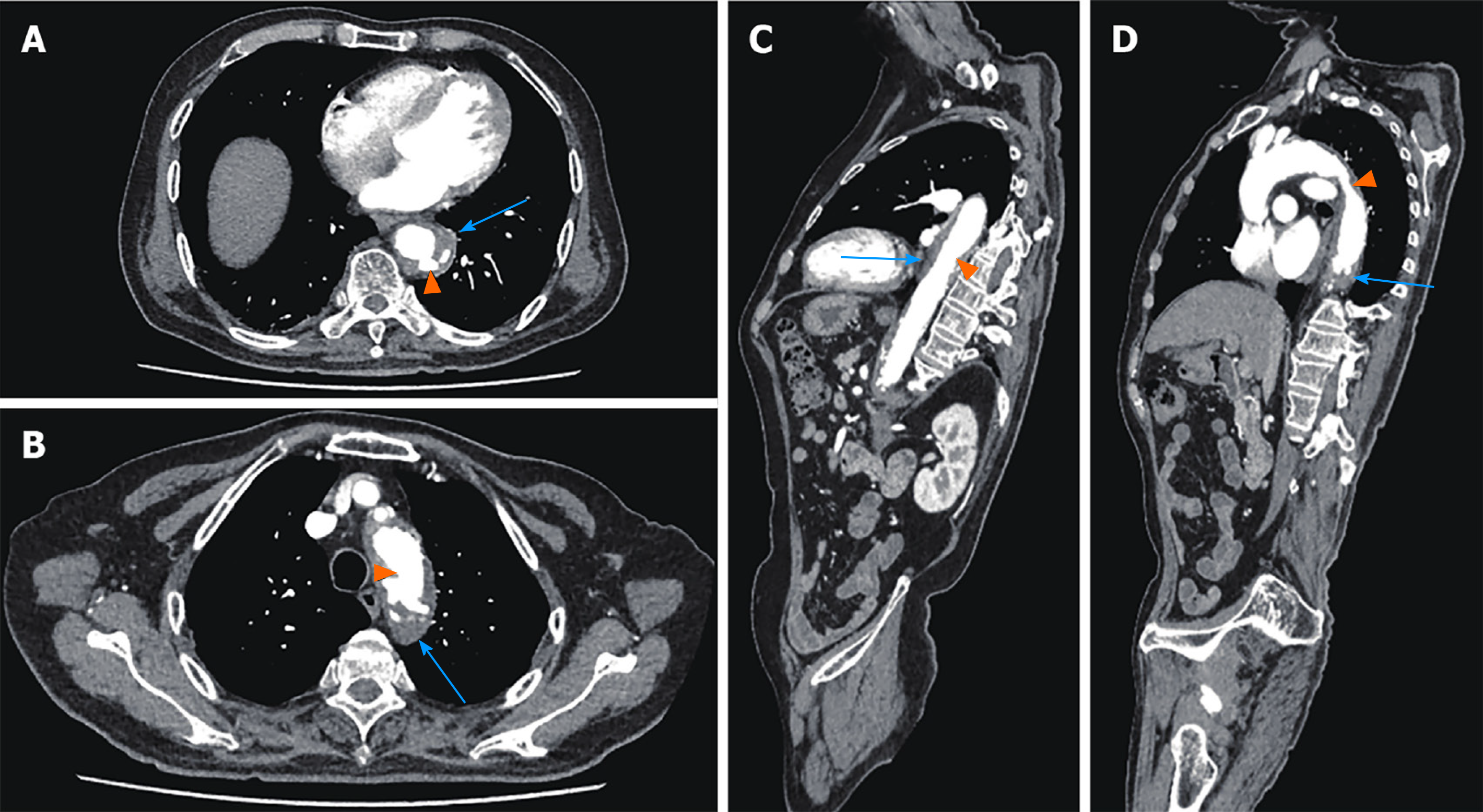
Figure 1 Preoperative thoracic and abdominal aortic computed tomography angiography.
A and B: Axial Computed tomography (CT) angiography showed low-density shadow peri-descending aorta, indicating intramural hemorrhage and hematoma (blue arrows), and multiple penetrating ulcers (orange arrows); C and D: Sagittal CT angiography showed low-density shadow peri-descending aorta, indicating intramural hemorrhage and hematoma (blue arrows), and multiple penetrating ulcers (orange arrows).
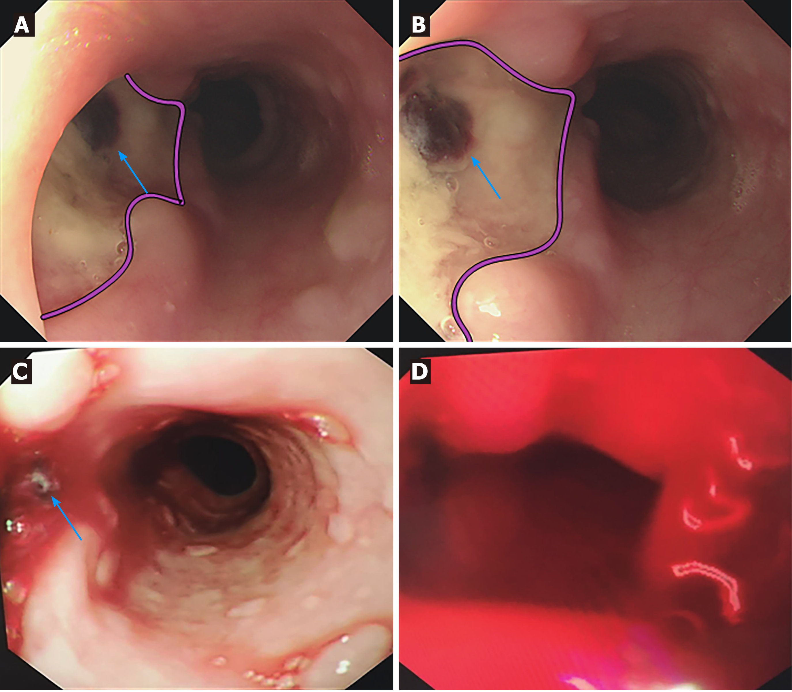
Figure 2 Esophageal endoscopic images of the patient.
A and B: First esophageal endoscopy showed a large deep longitudinal ulceration (2 cm × 1 cm) with an adherent clot (blue arrows) and white coating 25-27 cm below the incisors, corresponding to a fistula (pink lines showed the margin of the ulcer). No bleeding was found; C: Second bedside emergency endoscopy also showed blood clots (blue arrows); D: Second bedside emergency endoscopy showed massive bleeding as well.
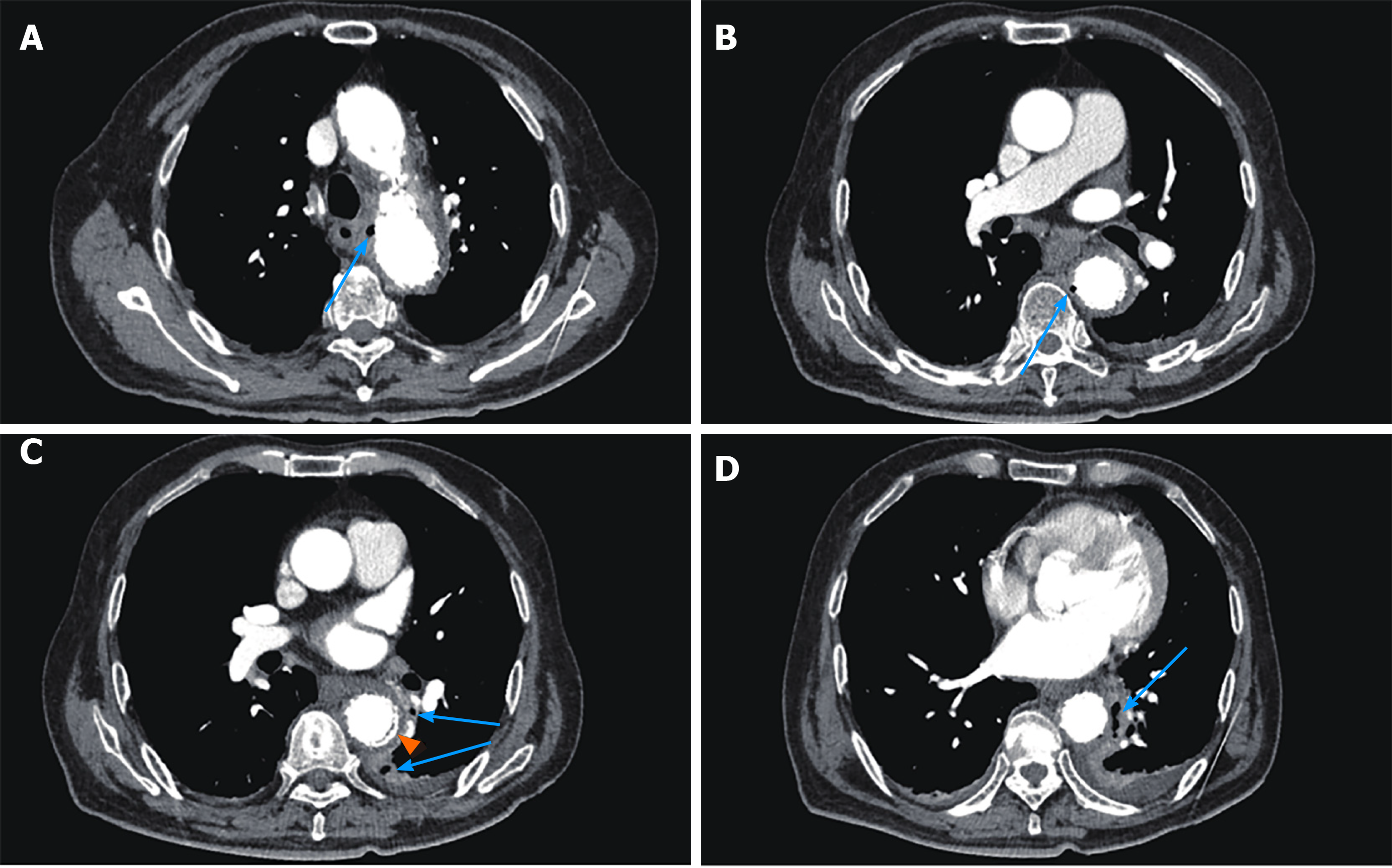
Figure 3 Postoperative thoracic and abdominal aortic computed tomography angiography.
A-D: Multiple occurrences of extraluminal air near the aortic stent graft (blue arrows); C: Intravenous contrast material outside the aortic stent graft (orange arrow).
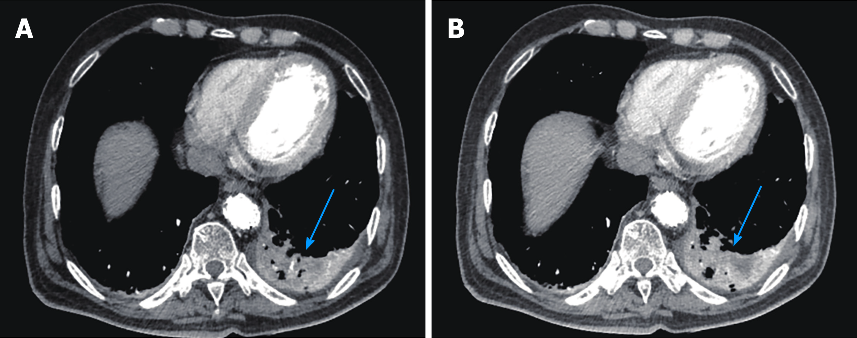
Figure 4 Pulmonary infection and empyema formation in postoperative thoracic and abdominal aortic computed tomography angiography.
Patchy soft tissue shadows adjacent to the aorta of the left lower lobe accompanied by cavity and empyema formation (blue arrows).
Figure 5 Aortoesophageal fistula formation in postoperative thoracic and abdominal aortic computed tomography angiography.
A and B: Rigid endovascular stent graft protruding to the esophageal lumen and in communication with the esophagus (blue arrows); C: Reconstructed image of the overall aorta and endovascular stent graft (blue arrows).
- Citation: Wang DQ, Liu M, Fan WJ. Secondary aortoesophageal fistula initially presented with empyema after thoracic aortic stent grafting: A case report. World J Clin Cases 2021; 9(29): 8938-8945
- URL: https://www.wjgnet.com/2307-8960/full/v9/i29/8938.htm
- DOI: https://dx.doi.org/10.12998/wjcc.v9.i29.8938


