Copyright
©The Author(s) 2021.
World J Clin Cases. Oct 16, 2021; 9(29): 8932-8937
Published online Oct 16, 2021. doi: 10.12998/wjcc.v9.i29.8932
Published online Oct 16, 2021. doi: 10.12998/wjcc.v9.i29.8932
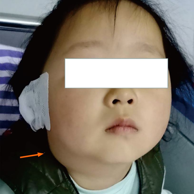
Figure 1
The patient’s right submaxillary and submental areas were swollen (marked by orange arrow).
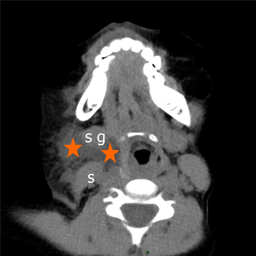
Figure 2 Neck computed tomography showed low density soft tissue shadow in the right submandibular space, laryngeal shift to the left, and subcutaneous tissue thickening.
Sternocleidomastoid (s); Submandibular gland (sg); Blood accumulation (orange star).
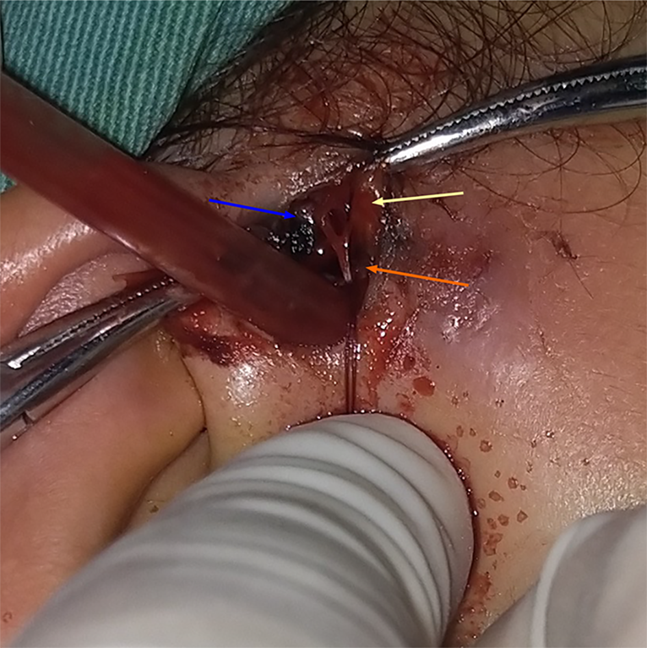
Figure 3 Intraoperative bleeding of the superficial temporal artery.
Superficial temporal artery (orange arrow); Superficial temporal artery, frontal branch (yellow arrow); Superficial temporal artery, parietal branch (blue arrow).
- Citation: Tian CH, Chen XJ. Severe bleeding after operation of preauricular fistula: A case report. World J Clin Cases 2021; 9(29): 8932-8937
- URL: https://www.wjgnet.com/2307-8960/full/v9/i29/8932.htm
- DOI: https://dx.doi.org/10.12998/wjcc.v9.i29.8932


