Copyright
©The Author(s) 2021.
World J Clin Cases. Oct 16, 2021; 9(29): 8923-8931
Published online Oct 16, 2021. doi: 10.12998/wjcc.v9.i29.8923
Published online Oct 16, 2021. doi: 10.12998/wjcc.v9.i29.8923
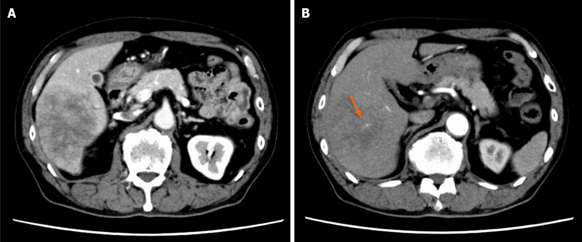
Figure 1 Image findings of contrast-enhanced dynamic-computed tomography.
A: Image showing a tumor with a diameter of approximately 8 cm in the posterior segment of the liver, which was weakly and gradually enhanced; B: Image showing an intratumoral artery in the arterial phase (arrow).
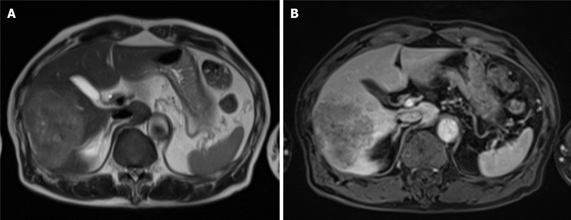
Figure 2 Image findings of magnetic resonance imaging.
A: The tumor showed high signal intensity on T2-weighted images; B: The tumor showed weak enhancement compared to surrounding liver parenchyma in the portal venous phase after administration of gadolinium ethoxybenzyl diethylenetriamine pentaacetic acid.
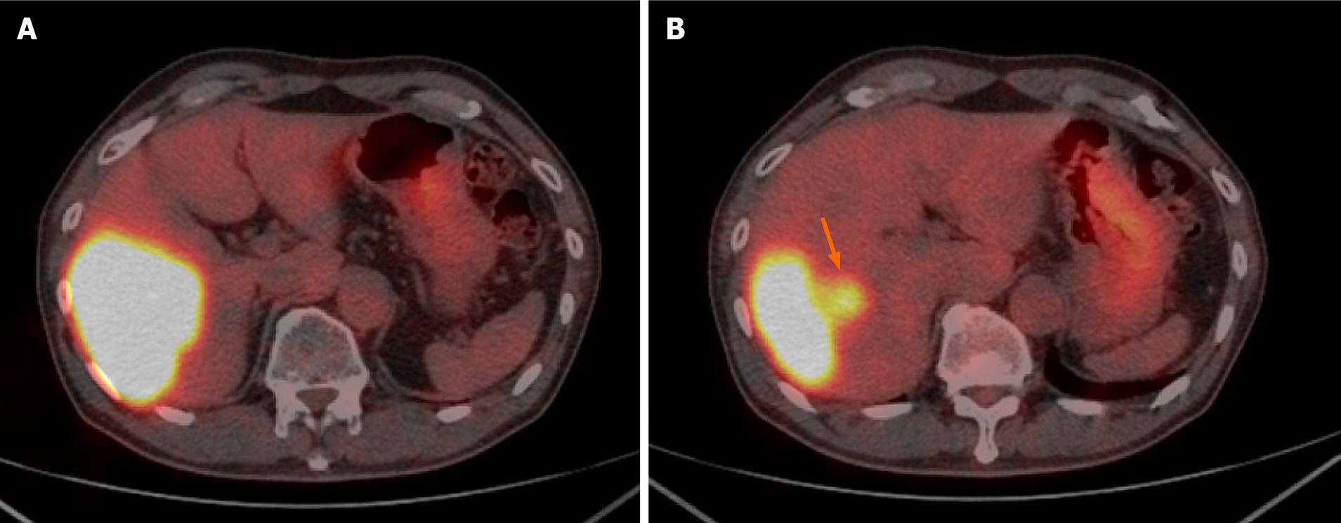
Figure 3 Image findings of 18F-fluorodeoxyglucose positron emission tomography/computed tomography.
A: Image showing an abnormally high uptake on the tumorous lesion in the posterior segment, with a maximum standardized uptake value (SUVmax) of 20.0; B: The abnormally high uptake showed that the tumor appeared to spread convexly along the intrahepatic bile ducts (arrow).
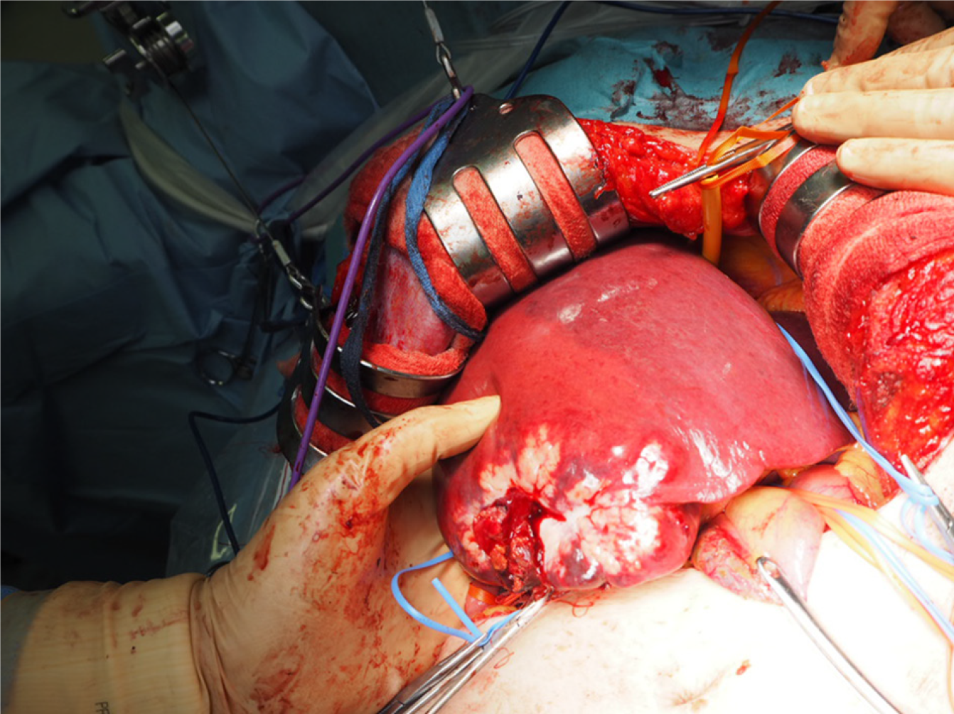
Figure 4 Intraoperative findings.
A large whitish tumor was partially exposed on the surface of the liver.
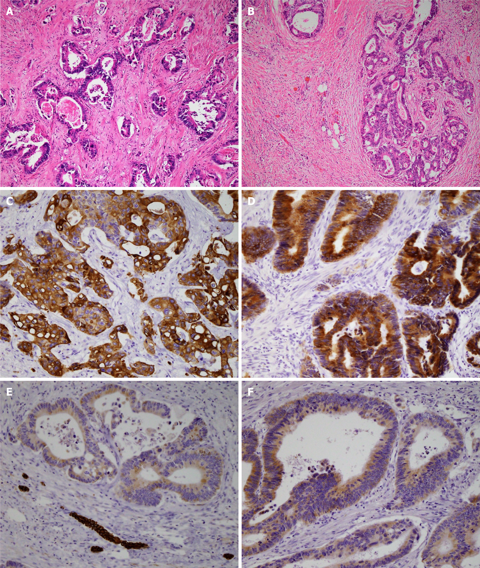
Figure 5 Histopathological findings.
A: The liver tumor represented a moderately differentiated adenocarcinoma with tubular and cribriform growth patterns (hematoxylin and eosin (H&E) staining at × 10 magnification); B: The rectal cancer represented a moderately differentiated adenocarcinoma (H&E staining at × 10 magnification). The image of the liver tumor showed that its pathological characteristics were the same as the primary rectal cancer; C: The liver tumor cells were positive for CK20 (immunostaining at × 20 magnification); D: The rectal cancer cells were positive for CK20 (immunostaining at × 20 magnification); E: The liver tumor cells were weakly positive for CK7 (immunostaining at × 20 magnification); F: The rectal cancer cells were weakly positive for CK7 (immunostaining at × 20 magnification).
- Citation: Yonenaga Y, Yokoyama S. Isolated liver metastasis detected 11 years after the curative resection of rectal cancer: A case report. World J Clin Cases 2021; 9(29): 8923-8931
- URL: https://www.wjgnet.com/2307-8960/full/v9/i29/8923.htm
- DOI: https://dx.doi.org/10.12998/wjcc.v9.i29.8923


