Copyright
©The Author(s) 2021.
World J Clin Cases. Oct 16, 2021; 9(29): 8915-8922
Published online Oct 16, 2021. doi: 10.12998/wjcc.v9.i29.8915
Published online Oct 16, 2021. doi: 10.12998/wjcc.v9.i29.8915
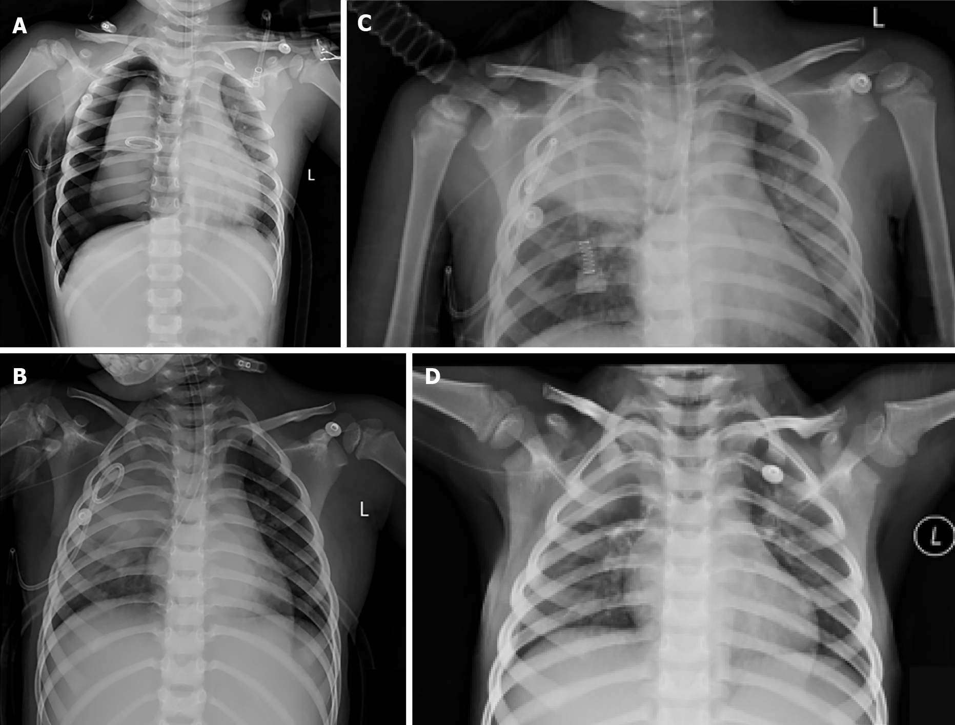
Figure 1 X-ray image.
A: Initial X-ray of the chest. After chest tube insertion for bilateral pneumothoraxes, massive air leak and failure of right lung expansion occurred due to right bronchial rupture. Fracture of the first and second anterior right rib, subcutaneous emphysema and right mediastinal hernia were observed; B: The 2nd X-ray of the chest. After ventilation using the modified tube, the air leak was stopped and there was spontaneous resolution of the pneumothorax, with limited subcutaneous emphysema. The end of the tracheal tube was located in the right main bronchus at the level of the lower margin of the 6th thoracic vertebra; C: Imaging on the 6th day. The right pneumothorax was significantly absorbed and the subcutaneous pneumatosis of the right chest wall was significantly decreased. There was less consolidation in both lungs than before; D: Follow-up X-ray 10 d after surgery. The transparency of the right upper lung field was still increased. The lesions in the middle and lower lung fields were basically absorbed, and the manifestations of the right pleural effusion were the same as before.
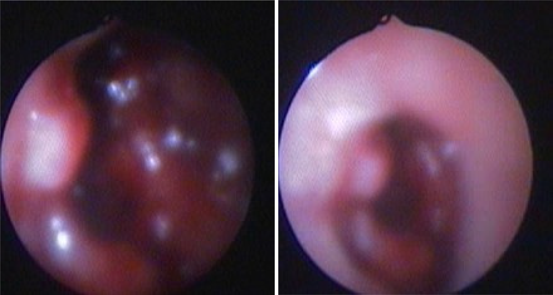
Figure 2
Hemorrhage due to rupture of the right upper lobe bronchus could be seen under fiberoptic bronchoscopy.
Figure 3
An incision was made in the tracheal tube.
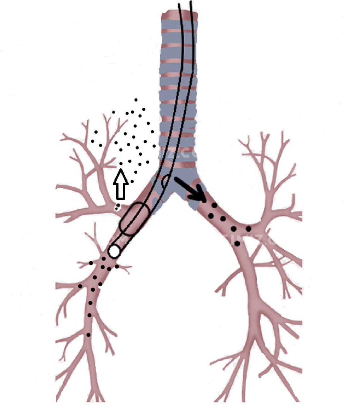
Figure 4 The modified tube was inserted under the guidance of a fiberoptic bronchoscope.
The incision in the tube was near the carina, which made it easy to ventilate the left lung through the incision. After the balloon was inflated, the balloon blocked the rupture in the bronchus. Using this method, the other right and left lung lobes were ventilated, thus avoiding atelectasis.
- Citation: Fan QM, Yang WG. Use of a modified tracheal tube in a child with traumatic bronchial rupture: A case report and review of literature. World J Clin Cases 2021; 9(29): 8915-8922
- URL: https://www.wjgnet.com/2307-8960/full/v9/i29/8915.htm
- DOI: https://dx.doi.org/10.12998/wjcc.v9.i29.8915


