Copyright
©The Author(s) 2021.
World J Clin Cases. Oct 16, 2021; 9(29): 8831-8838
Published online Oct 16, 2021. doi: 10.12998/wjcc.v9.i29.8831
Published online Oct 16, 2021. doi: 10.12998/wjcc.v9.i29.8831
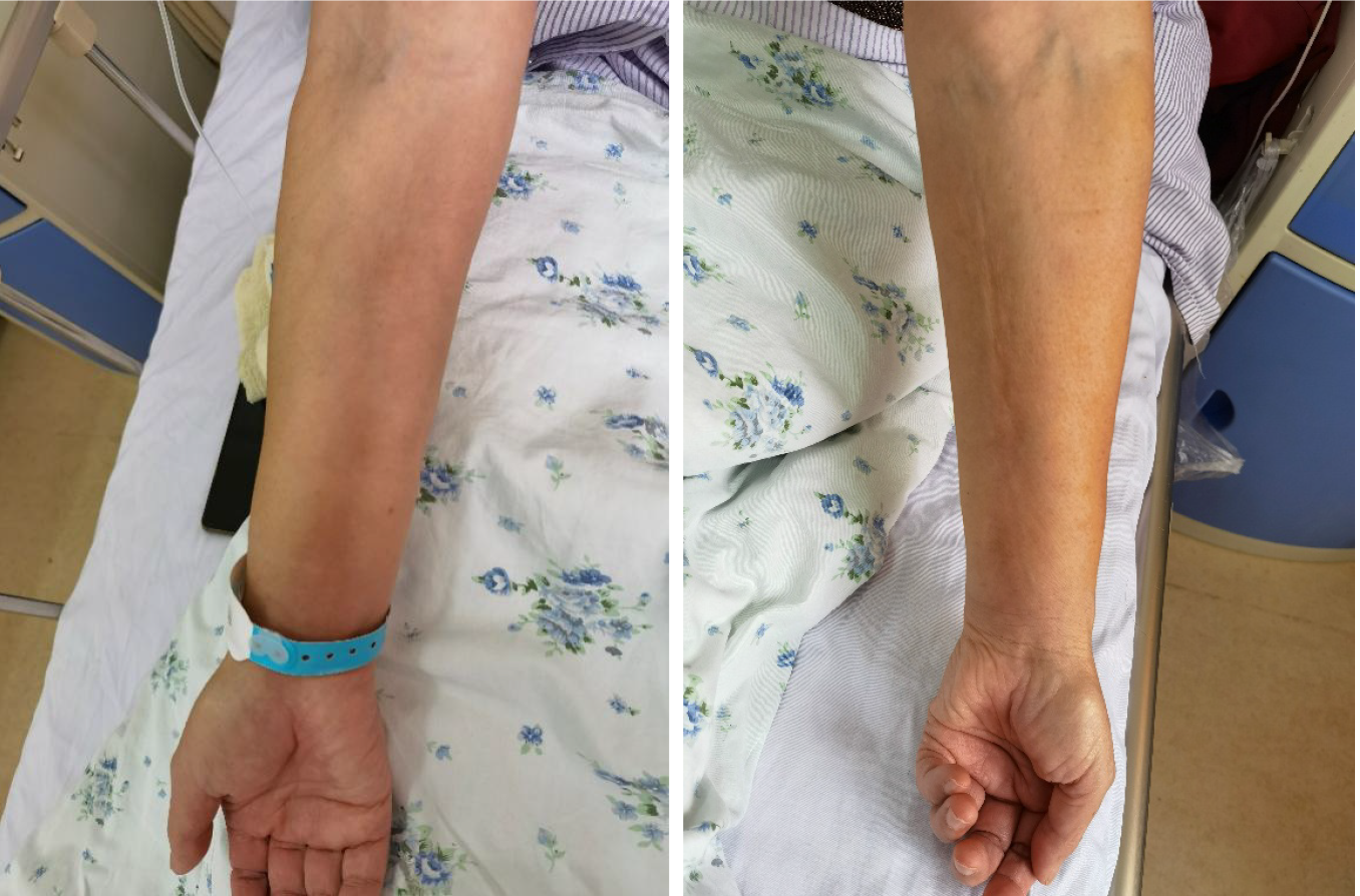
Figure 1 The skin and muscle of Case 1’s upper limbs were stiff, tight, and smooth with pitting.
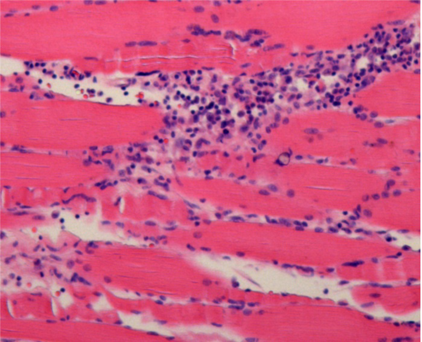
Figure 2 Histopathological examination.
The paraffin section specimen was taken from the left upper arm muscle, stained with histopathological examination, and examined at magnification (100 ×). We observed epidermal atrophy and thinning, deep subcutaneous fascia fiber proliferation, and considerable infiltration by chronic inflammatory cells and eosinophils. Fibers of the perineurium and intermuscular membrane had proliferated, and striated muscle cells at the junction of the sarcolemma had been destroyed.
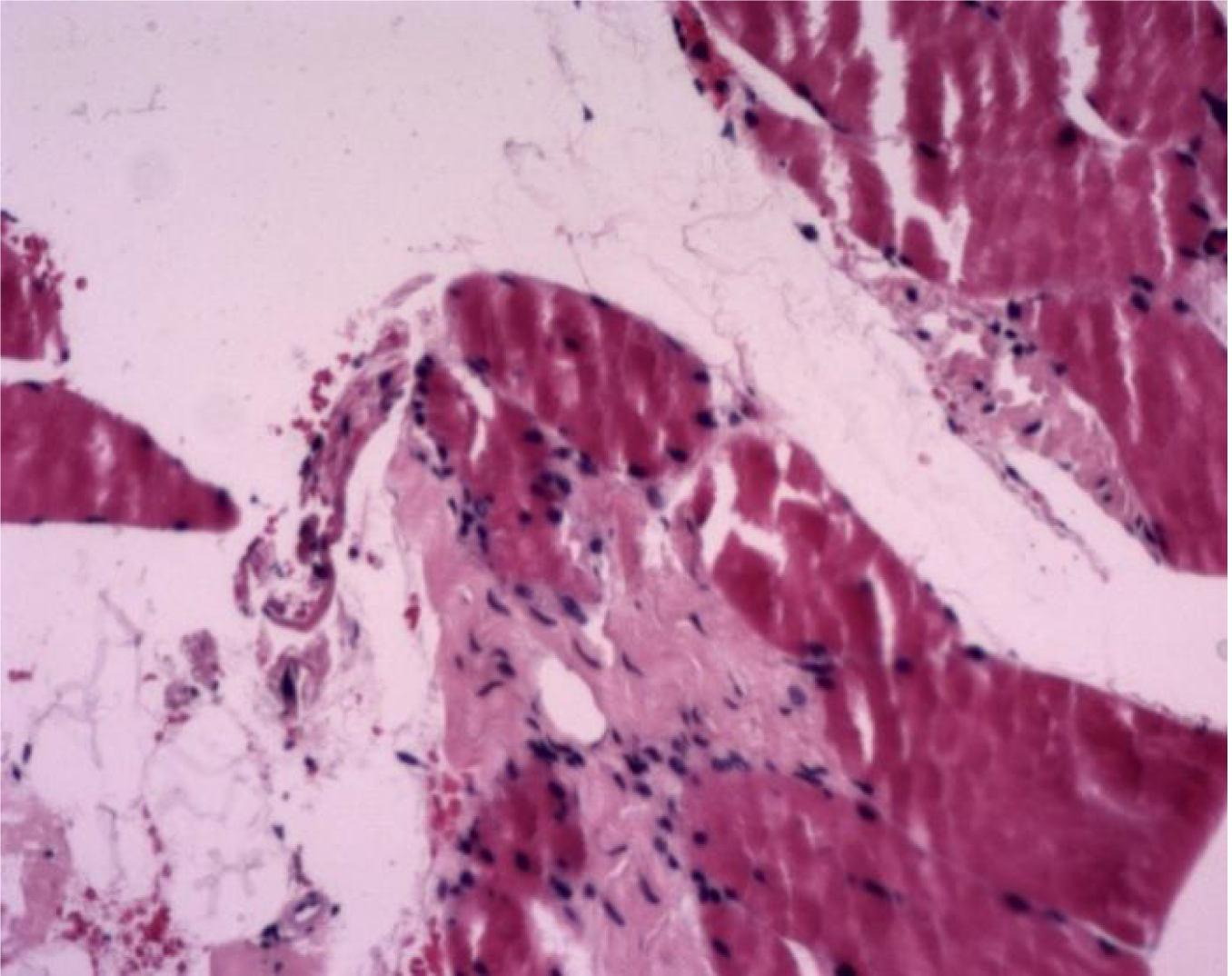
Figure 3 Histopathological examination.
Paraffin section specimens were taken from the right lower limb muscles, stained with histopathological examination, and examined at magnification (100 ×). Skeletal muscles were visible in the broken tissues, some of the horizontal stripes were unclear, and there was proliferation of fibrous tissue and small blood vessels.
- Citation: Song Y, Zhang N, Yu Y. Diagnosis and treatment of eosinophilic fasciitis: Report of two cases. World J Clin Cases 2021; 9(29): 8831-8838
- URL: https://www.wjgnet.com/2307-8960/full/v9/i29/8831.htm
- DOI: https://dx.doi.org/10.12998/wjcc.v9.i29.8831


