Copyright
©The Author(s) 2021.
World J Clin Cases. Oct 16, 2021; 9(29): 8820-8824
Published online Oct 16, 2021. doi: 10.12998/wjcc.v9.i29.8820
Published online Oct 16, 2021. doi: 10.12998/wjcc.v9.i29.8820
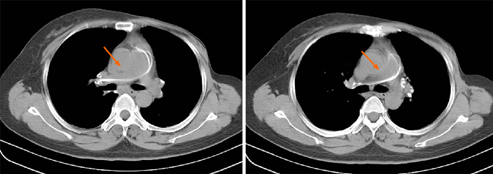
Figure 1 Computed tomography.
Both axial image showing a strip high density shadow in the main pulmonary artery-right pulmonary artery-hilar area (orange arrows).
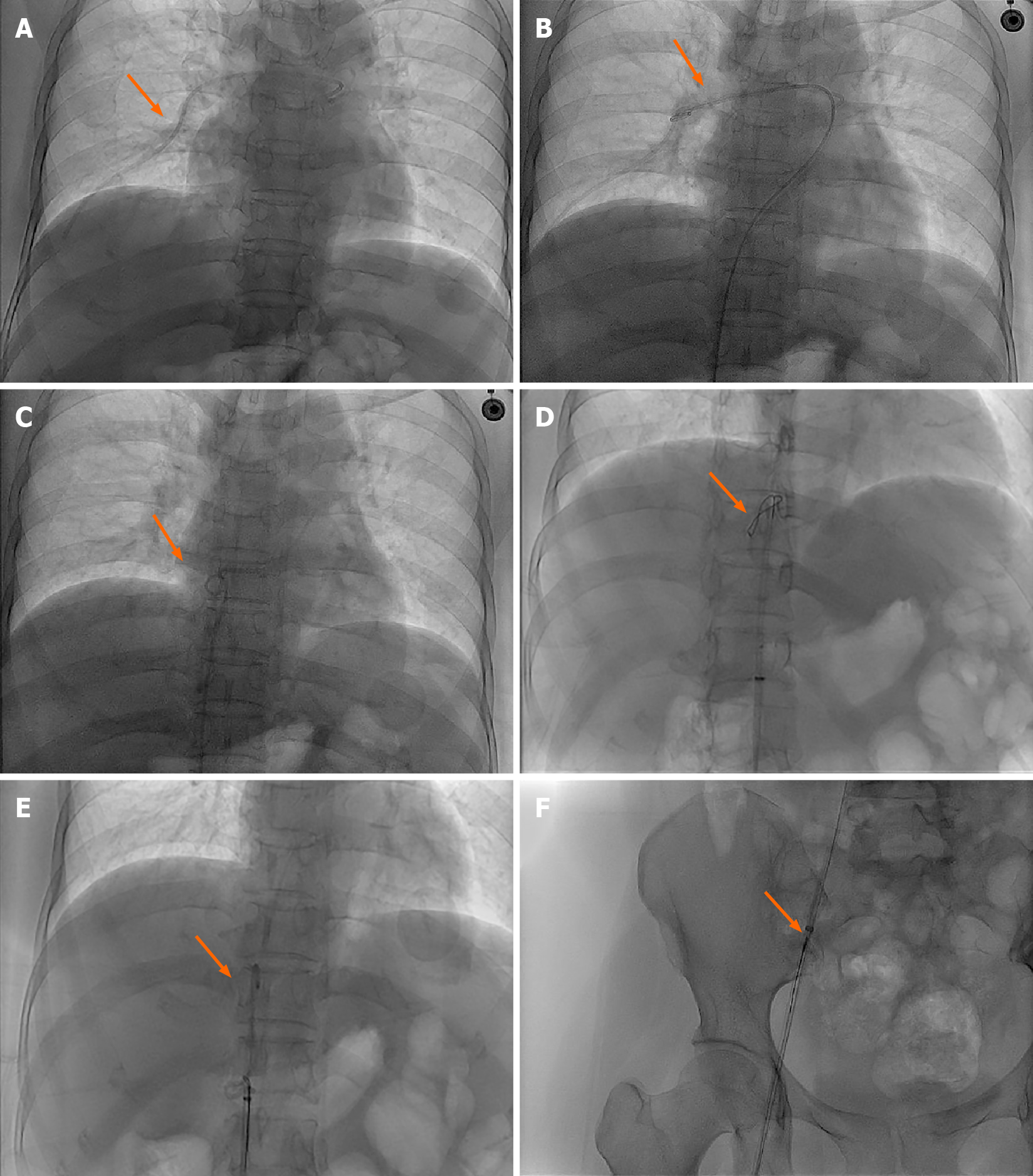
Figure 2 Digital subtraction angiography images.
A: The main pulmonary artery and right pulmonary artery showed tubular shadow (orange arrow); B-F: A 5F pigtail catheter was selected, and the dislodged catheter was towed to the inferior vena cava after the two catheters were crossed by rotating the catheter outside the body according to the natural bending at the front end of the catheter, The intravenous infusion port catheter was then successfully removed using a 5F gooseneck trap (orange arrows).
- Citation: Chen GQ, Wu Y, Zhao KF, Shi RS. Removal of "ruptured" pulmonary artery infusion port catheter by pigtail catheter combined with gooseneck trap: A case report. World J Clin Cases 2021; 9(29): 8820-8824
- URL: https://www.wjgnet.com/2307-8960/full/v9/i29/8820.htm
- DOI: https://dx.doi.org/10.12998/wjcc.v9.i29.8820


