Copyright
©The Author(s) 2021.
World J Clin Cases. Oct 16, 2021; 9(29): 8804-8811
Published online Oct 16, 2021. doi: 10.12998/wjcc.v9.i29.8804
Published online Oct 16, 2021. doi: 10.12998/wjcc.v9.i29.8804
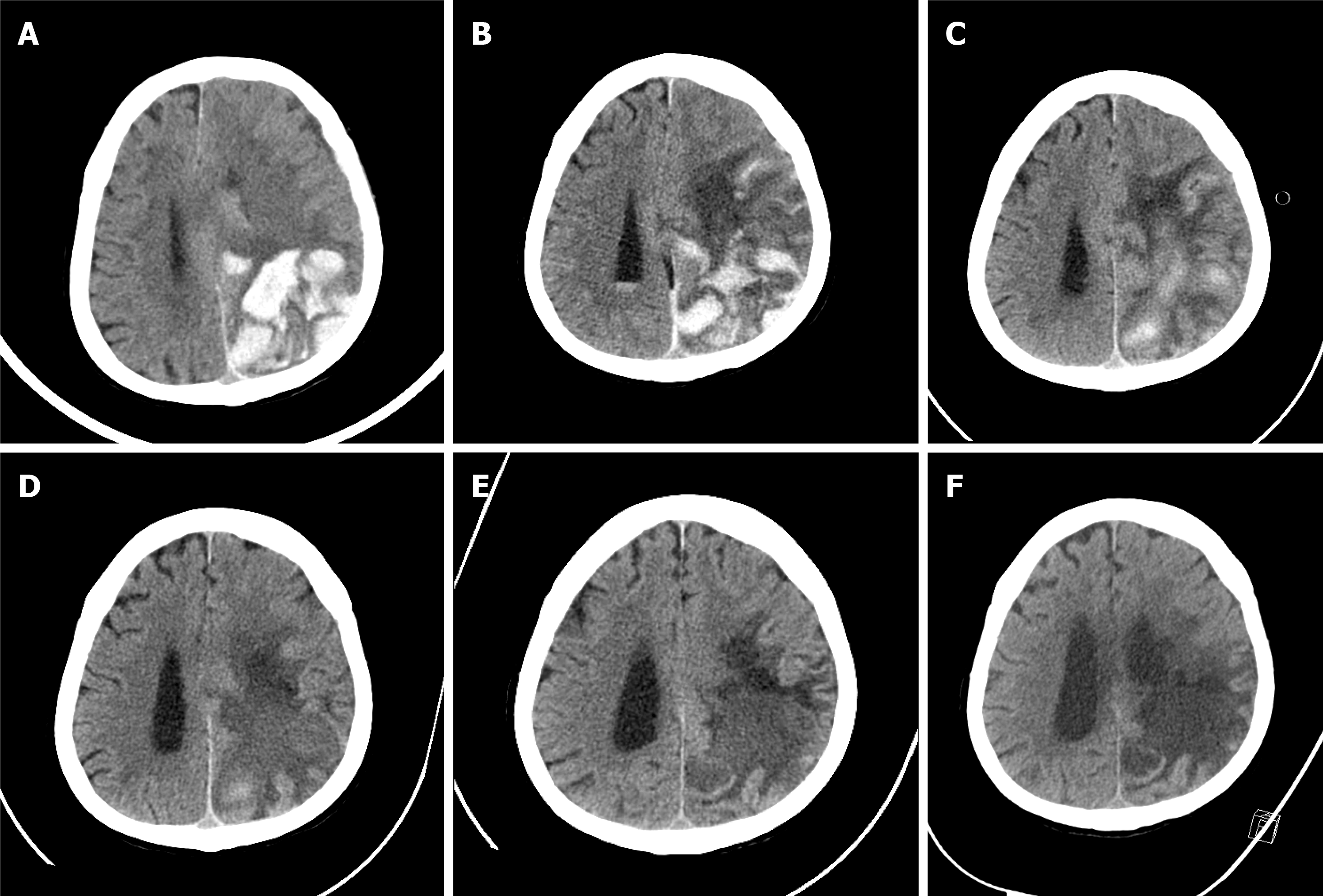
Figure 1 Change in brain computed tomography during hospitalization.
A: Brain computed tomography (CT) at admission. Intracranial hemorrhage in the parieto-occipital lobe was confirmed, and midline shift was observed due to brain edema; B: Brain CT on the 8th hospital day. Intracranial hemorrhage persists and concomitant Intra-ventricular hemorrhage (IVH) is confirmed; C: Brain CT on the 17th hospital day. Intracranial hemorrhage has begun to resolve and improvement of IVH is shown; D: Brain CT on the 28th hospital day. Intracranial hemorrhage shows ongoing resolution, and brain edema has also decreased; E: Brain CT on the 57th hospital day. Improvement in brain edema has resulted in dilatation of the ventricle and resolution of midline shift; F: Brain CT on the 82nd hospital day. Encephalomalacic change in the left parietal lobe is confirmed, and there is no evidence of new intracranial hemorrhage.
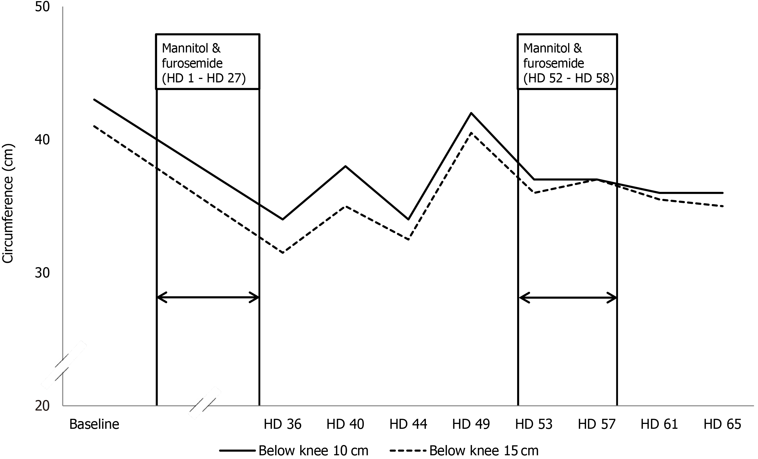
Figure 2 Change of circumference of the left lower leg before and after administration of mannitol and furosemide.
After the patient received mannitol and furosemide from the 1st to the 27th hospital day, the left lower extremity lymphedema improved dramatically. The improved lymphedema was also identified after re-administration of mannitol and furosemide from the 52th to the 58th hospital day. HD: Hospital day.
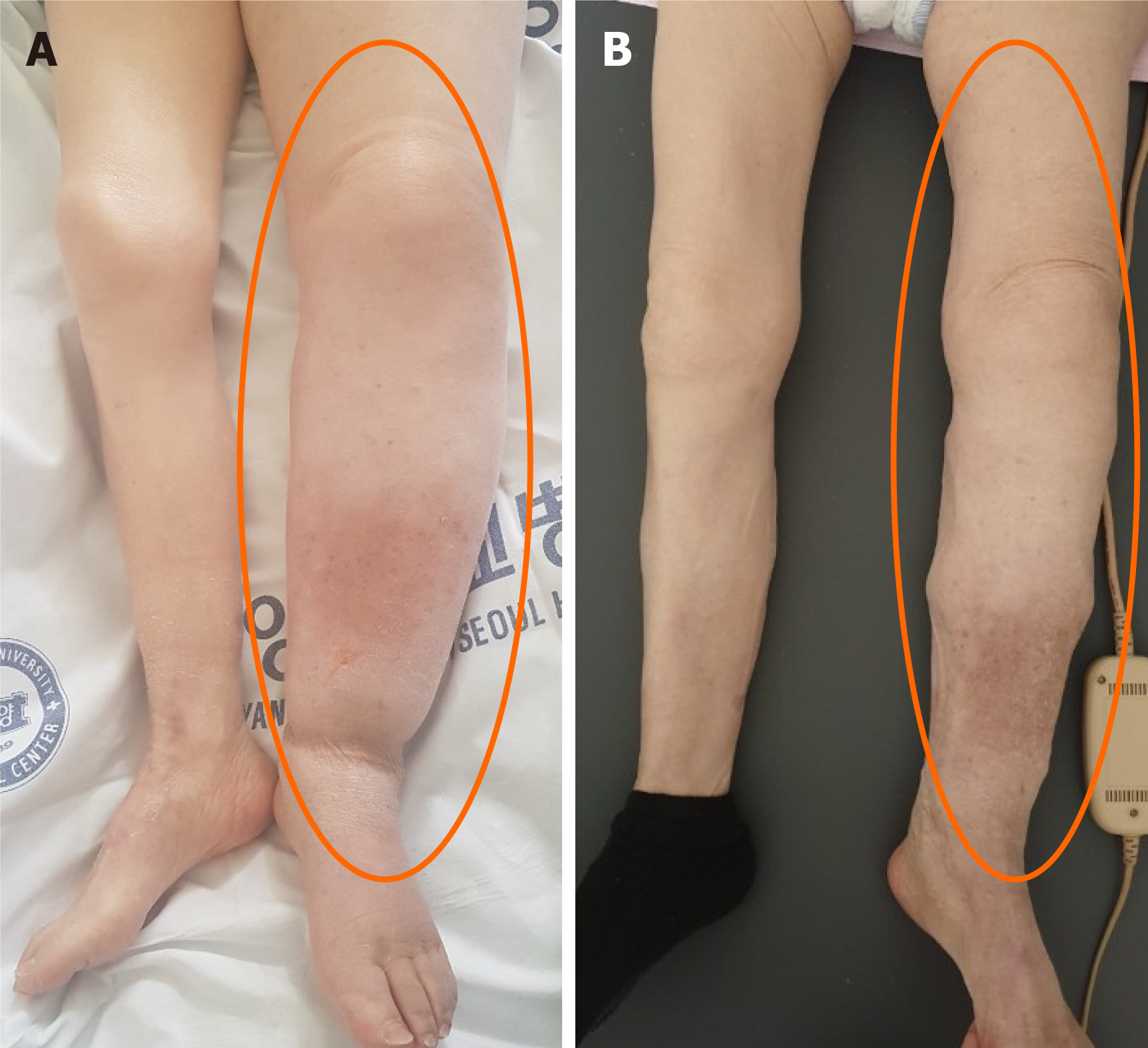
Figure 3 Changes in lymphedema during the hospital day.
A: After discontinuation of mannitol and furosemide (on the 49th hospital day); B: Improved lymphedema after reinstatement of mannitol and furosemide (on the 61st hospital day).
- Citation: Kim HS, Lee JY, Jung JW, Lee KH, Kim MJ, Park SB. Is mannitol combined with furosemide a new treatment for refractory lymphedema? A case report. World J Clin Cases 2021; 9(29): 8804-8811
- URL: https://www.wjgnet.com/2307-8960/full/v9/i29/8804.htm
- DOI: https://dx.doi.org/10.12998/wjcc.v9.i29.8804


