Copyright
©The Author(s) 2021.
World J Clin Cases. Oct 6, 2021; 9(28): 8563-8570
Published online Oct 6, 2021. doi: 10.12998/wjcc.v9.i28.8563
Published online Oct 6, 2021. doi: 10.12998/wjcc.v9.i28.8563
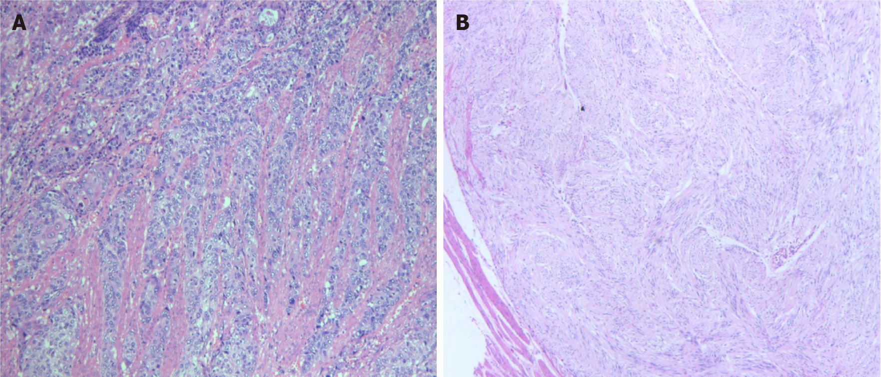
Figure 1 Postoperative histopathology of two lesions.
A: Postoperative histopathology of esophageal squamous-cell carcinoma lesion (magnifications, × 400); B: Postoperative histopathology of gastrointestinal stromal tumour lesion (magnifications, × 400).
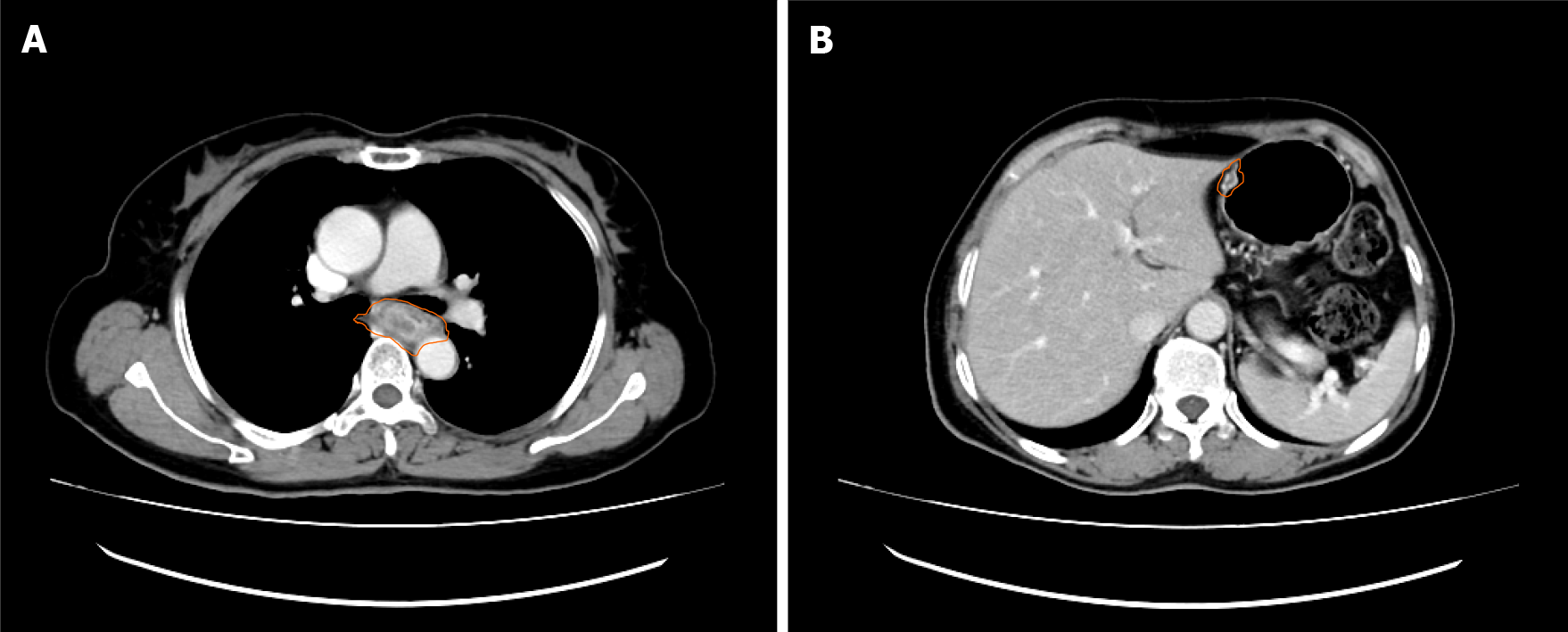
Figure 2 Preoperative computed tomography images.
A: Computed tomography (CT) scans of the chest showed a tumour in the esophagus; B: CT image of nodules of the lesser curvature of the stomach. The orange circles indicate tumour lesions.
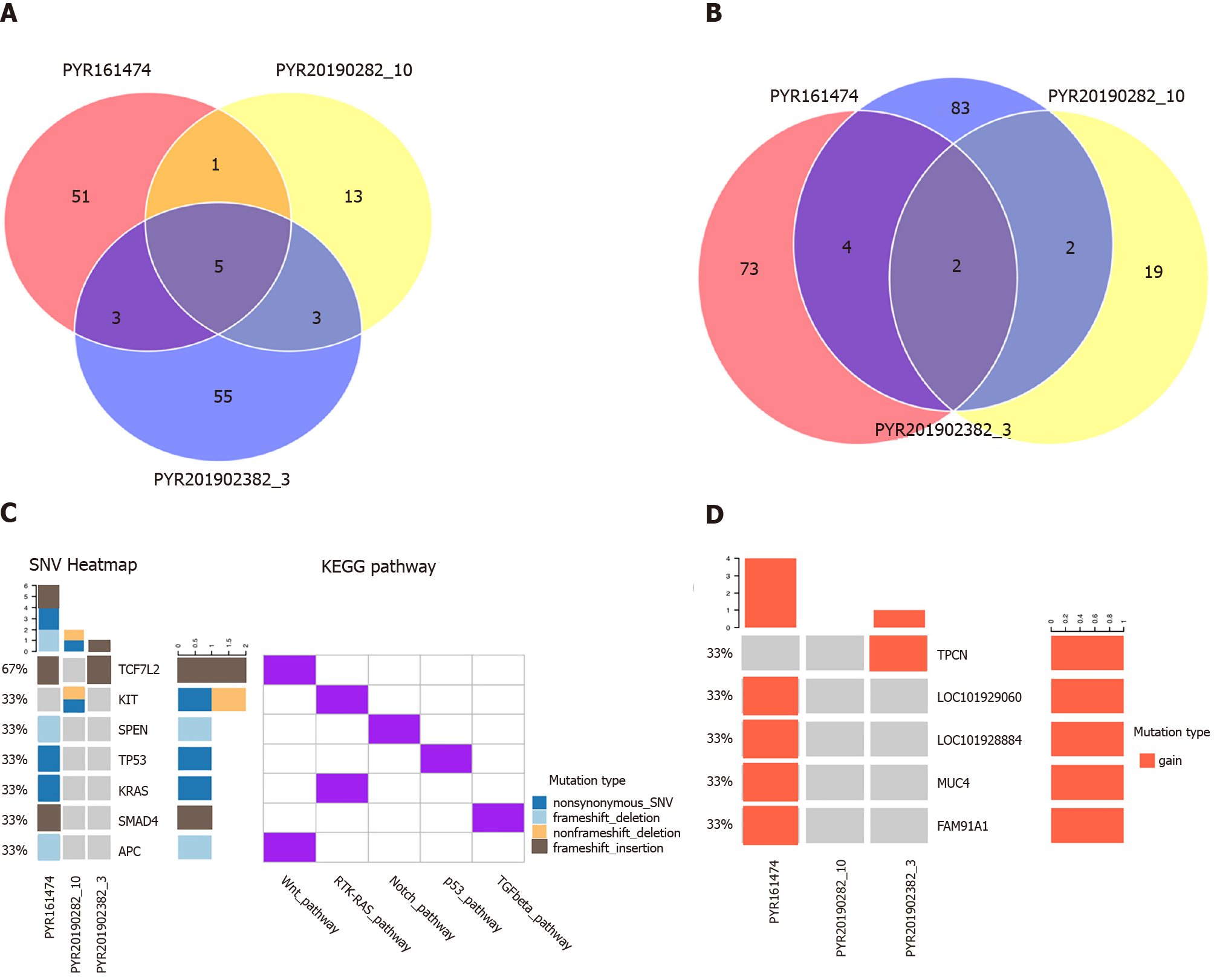
Figure 3 Genetic test results of three lesions.
A: Venn diagram of mutant genes; B: Venn diagram of mutant gene loci; C: Patterns of common gene variants and signaling pathways; D: Copy number variation heat map. PYP161474 represents tissue specimen of colorectal adenocarcinoma, PYP20190282-10 represents tissue specimen of gastrointestinal stromal tumour, and PYP201902382-3 represents tissue specimen of esophageal cancer.
- Citation: Ouyang WW, Li QY, Yang WG, Su SF, Wu LJ, Yang Y, Lu B. Genetic characteristics of a patient with multiple primary cancers: A case report. World J Clin Cases 2021; 9(28): 8563-8570
- URL: https://www.wjgnet.com/2307-8960/full/v9/i28/8563.htm
- DOI: https://dx.doi.org/10.12998/wjcc.v9.i28.8563


