Copyright
©The Author(s) 2021.
World J Clin Cases. Oct 6, 2021; 9(28): 8492-8497
Published online Oct 6, 2021. doi: 10.12998/wjcc.v9.i28.8492
Published online Oct 6, 2021. doi: 10.12998/wjcc.v9.i28.8492
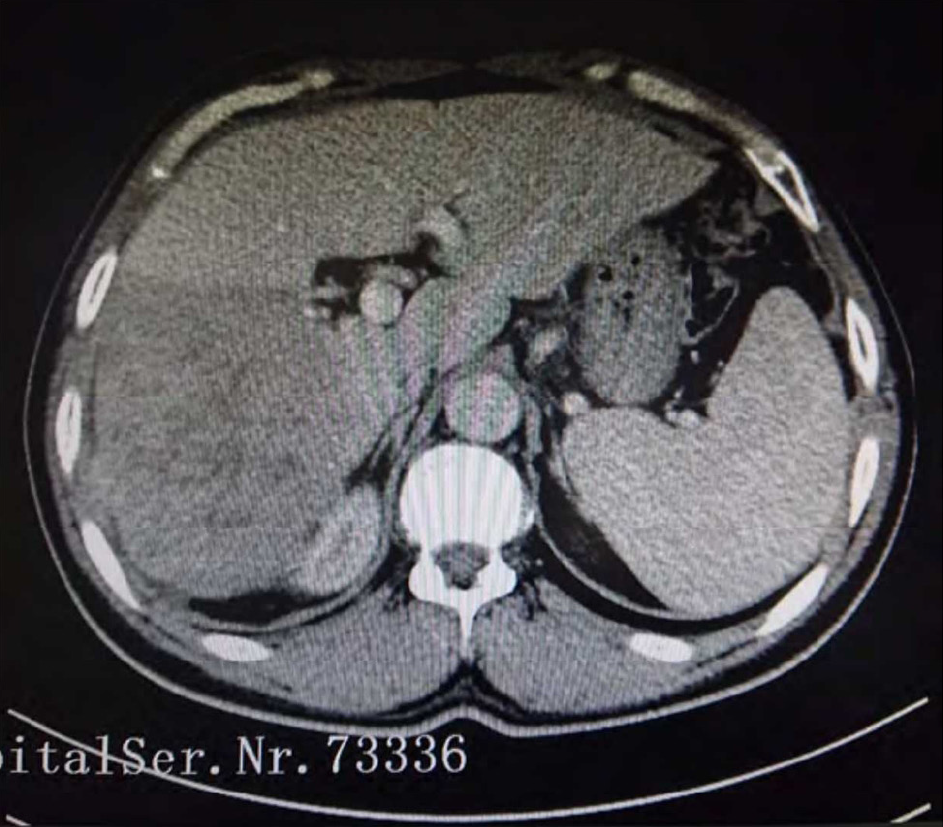
Figure 1 Representative computed tomography image used for hepatocellular carcinoma diagnosis.
A low-density soft tissue area was observed in the scanning plane of the upper abdomen. Mild density enhancement in the arterial phase and non-homogeneous density enhancement in the portal phase were observed. The tumor was about 10 cm × 12 cm in cross-section.
- Citation: Han JJ, Chen Y, Nan YC, Yang YL. Extremely high titer of hepatitis B surface antigen antibodies in a primary hepatocellular carcinoma patient: A case report. World J Clin Cases 2021; 9(28): 8492-8497
- URL: https://www.wjgnet.com/2307-8960/full/v9/i28/8492.htm
- DOI: https://dx.doi.org/10.12998/wjcc.v9.i28.8492


