Copyright
©The Author(s) 2021.
World J Clin Cases. Sep 26, 2021; 9(27): 8207-8213
Published online Sep 26, 2021. doi: 10.12998/wjcc.v9.i27.8207
Published online Sep 26, 2021. doi: 10.12998/wjcc.v9.i27.8207
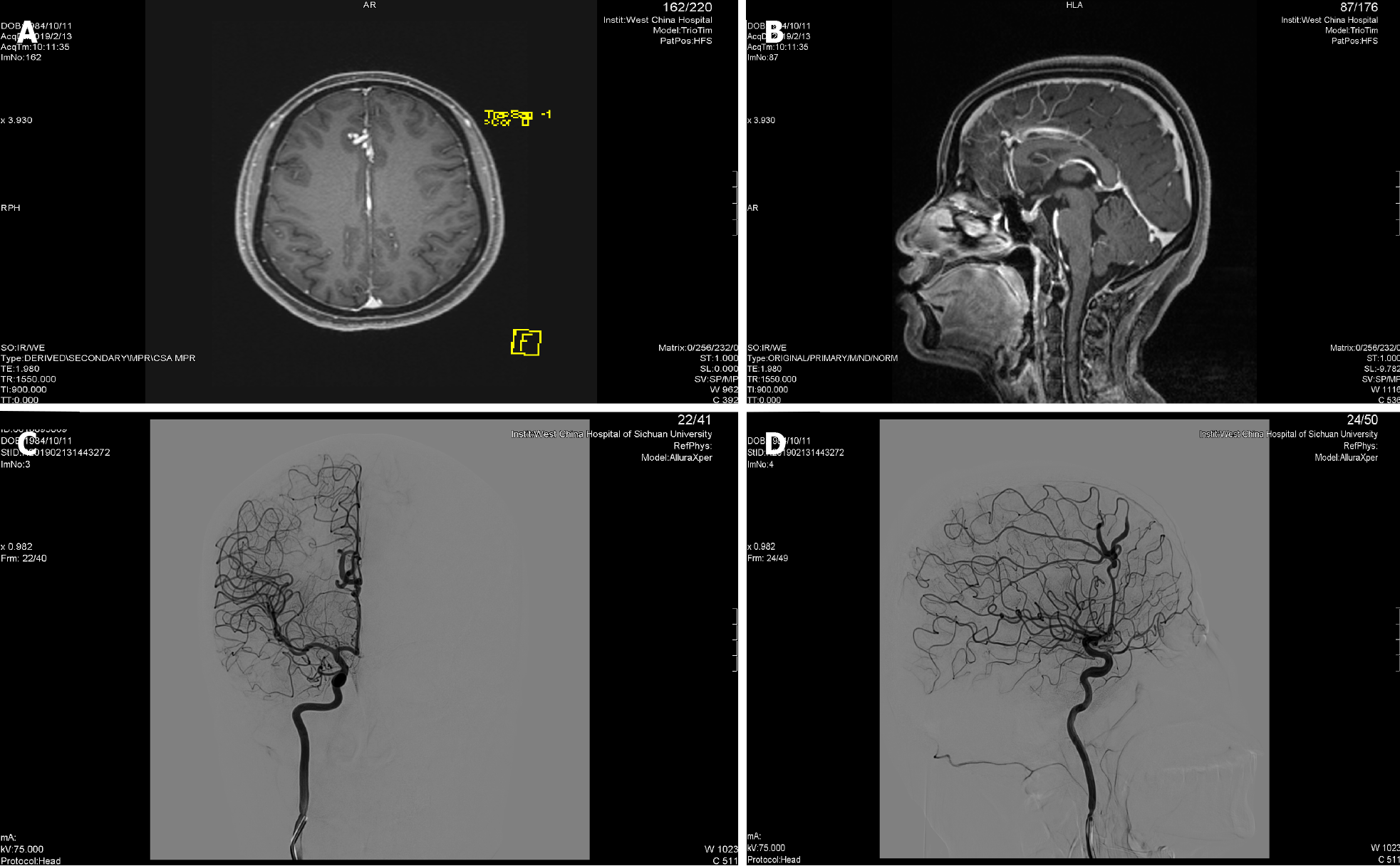
Figure 1 The preoperative magnetic resonance imaging of head showing high density shade in right front lobe and next to cerebral falx.
A: The axial magnetic resonance imaging (MRI); B: The sagittal MRI. The preoperative right internal carotid artery injection angiograms showing A3 segment malformation of the right anterior cerebral artery; C: Anteroposterior projection; D: Lateral projection.
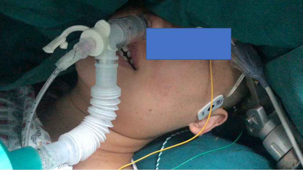
Figure 2 Intraoperative photograph showing the patient positioned in supine position with head rotated to the left.
She placed a nasopharyngeal airway.
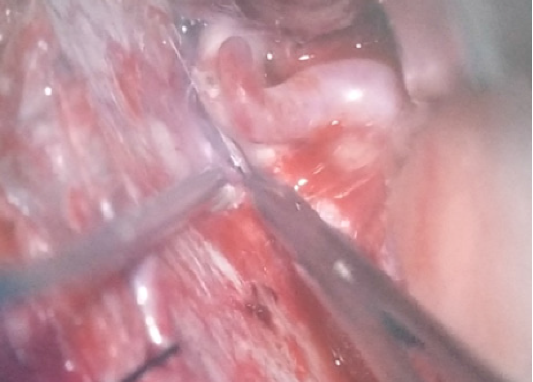
Figure 3 The intraoperative finding: The dilated and tortuous segment of the right anterior cerebral artery.
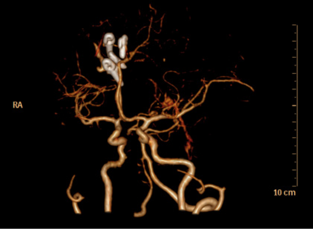
Figure 4 Postoperative computed tomography angiograph image: 3 clips used for aneurysm occlusion.
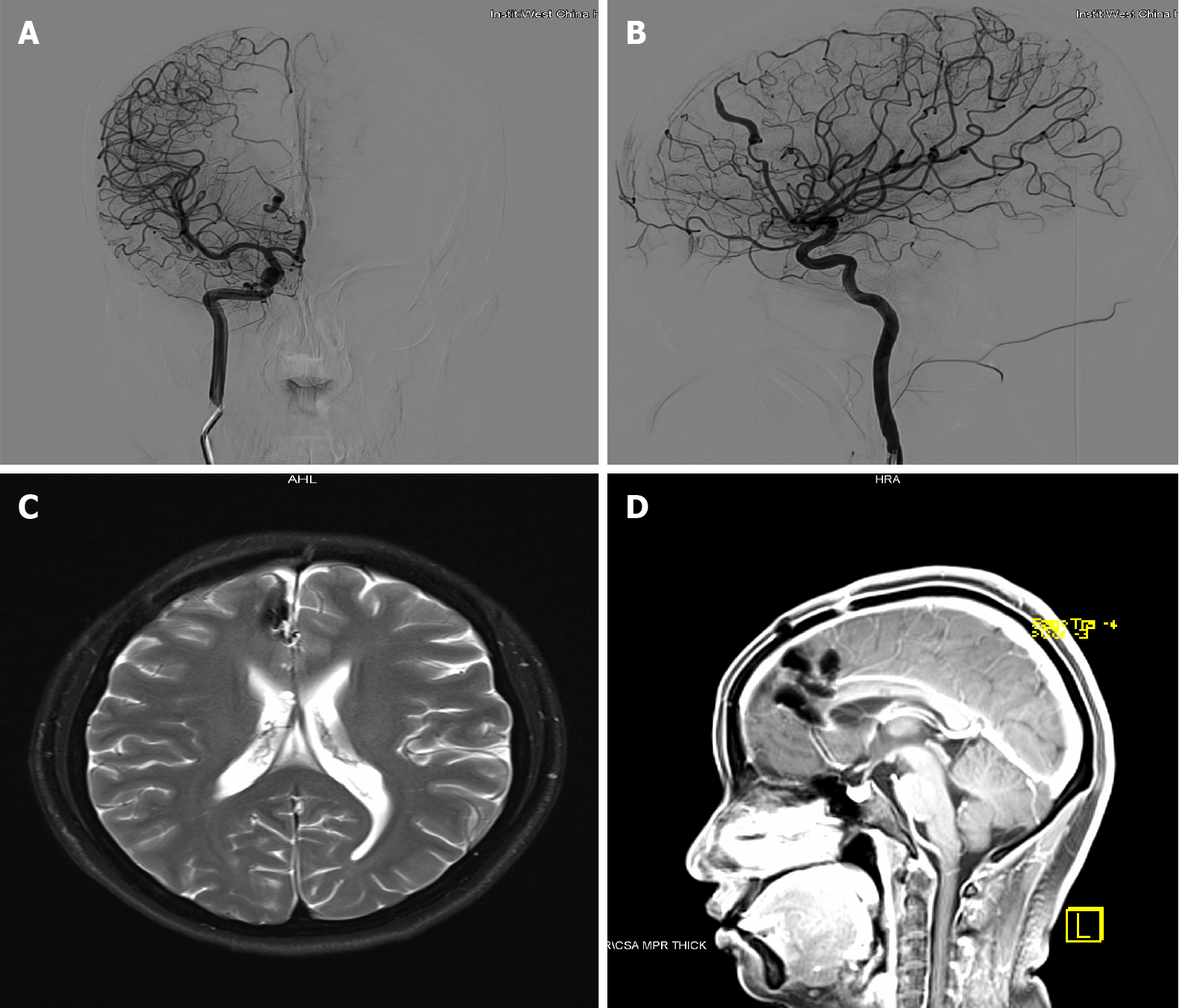
Figure 5 The postoperative right internal carotid artery injection angiograms showing no dilated and tortuous segment.
A: Anteroposterior projection; B: Lateral projection. The preoperative magnetic resonance imaging (MRI) of head showing no ischemic lesions; C: The axial MRI; D: The sagittal MRI.
- Citation: Zhou HY, Chen HY, Li Y. Anesthetic technique for awake artery malformation clipping with motor evoked potential and somatosensory evoked potential: A case report. World J Clin Cases 2021; 9(27): 8207-8213
- URL: https://www.wjgnet.com/2307-8960/full/v9/i27/8207.htm
- DOI: https://dx.doi.org/10.12998/wjcc.v9.i27.8207


