Copyright
©The Author(s) 2021.
World J Clin Cases. Sep 26, 2021; 9(27): 8147-8156
Published online Sep 26, 2021. doi: 10.12998/wjcc.v9.i27.8147
Published online Sep 26, 2021. doi: 10.12998/wjcc.v9.i27.8147
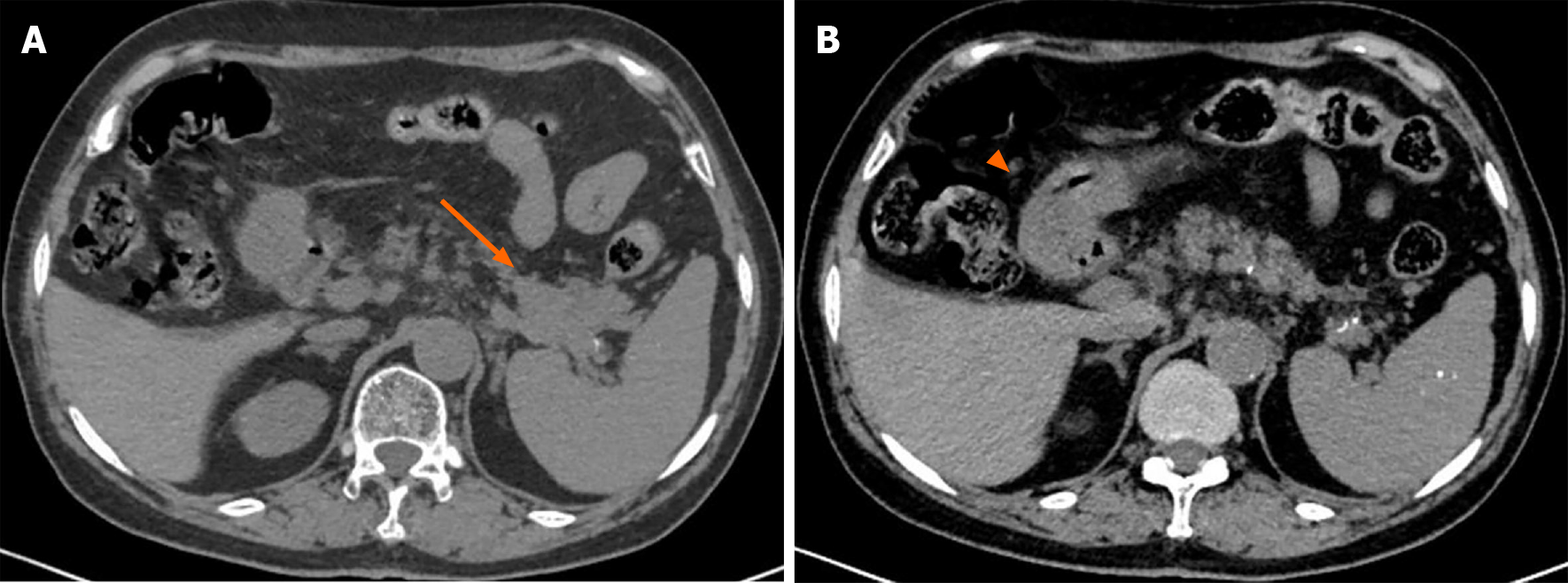
Figure 1 Upper abdominal computed tomography images.
A: A lobulated solid mass (orange arrow) in the cauda pancreas invading the splenic pedicle; B: The gastric antrum is full of chyme, and the accurate condition of the gastric wall is not clear (orange arrowhead).
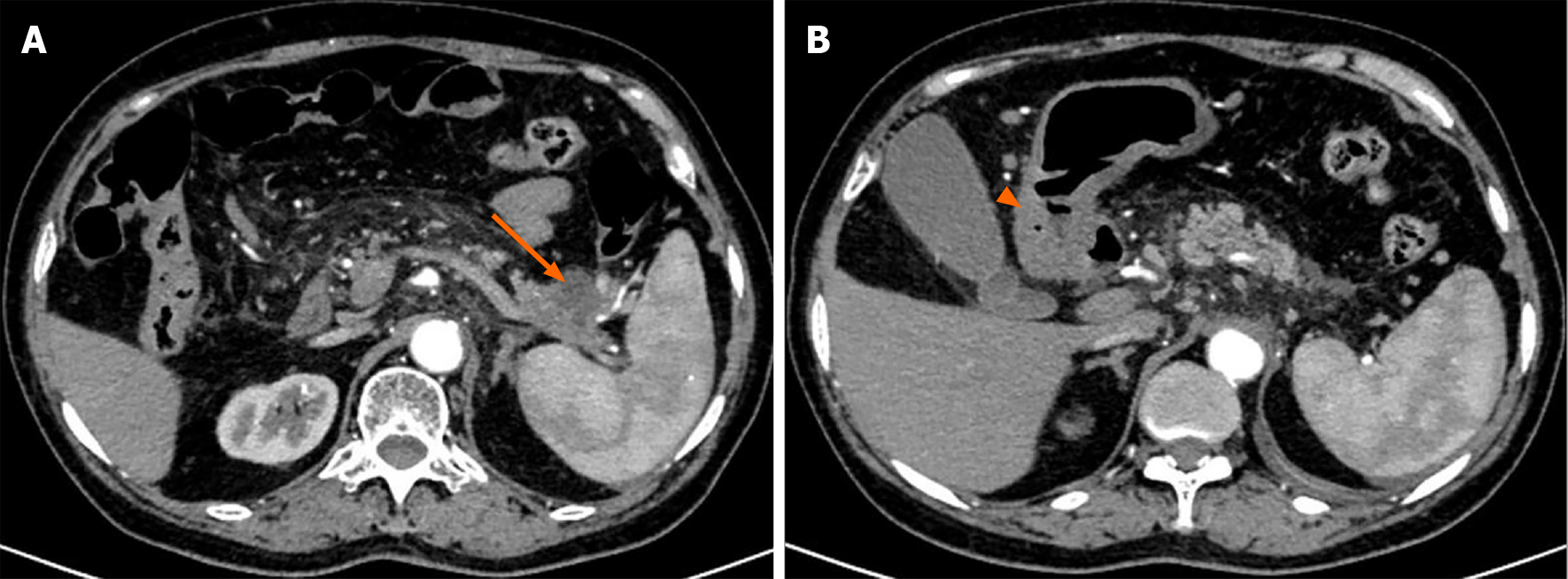
Figure 2 Enhanced upper abdominal computed tomography images.
A: A cystic solid mass measuring 3.5 cm × 3 cm × 2 cm in the cauda pancreas (orange arrow) surrounding the splenic vessels; B: Thickening of the antral wall and slight obstruction of the pylorus (orange arrowhead).
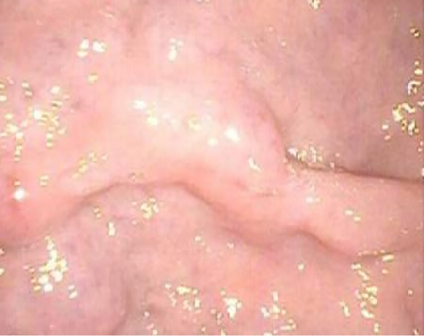
Figure 3 Gastric endoscopy revealed a submucosal lesion that arose from the surface of the pylorus.
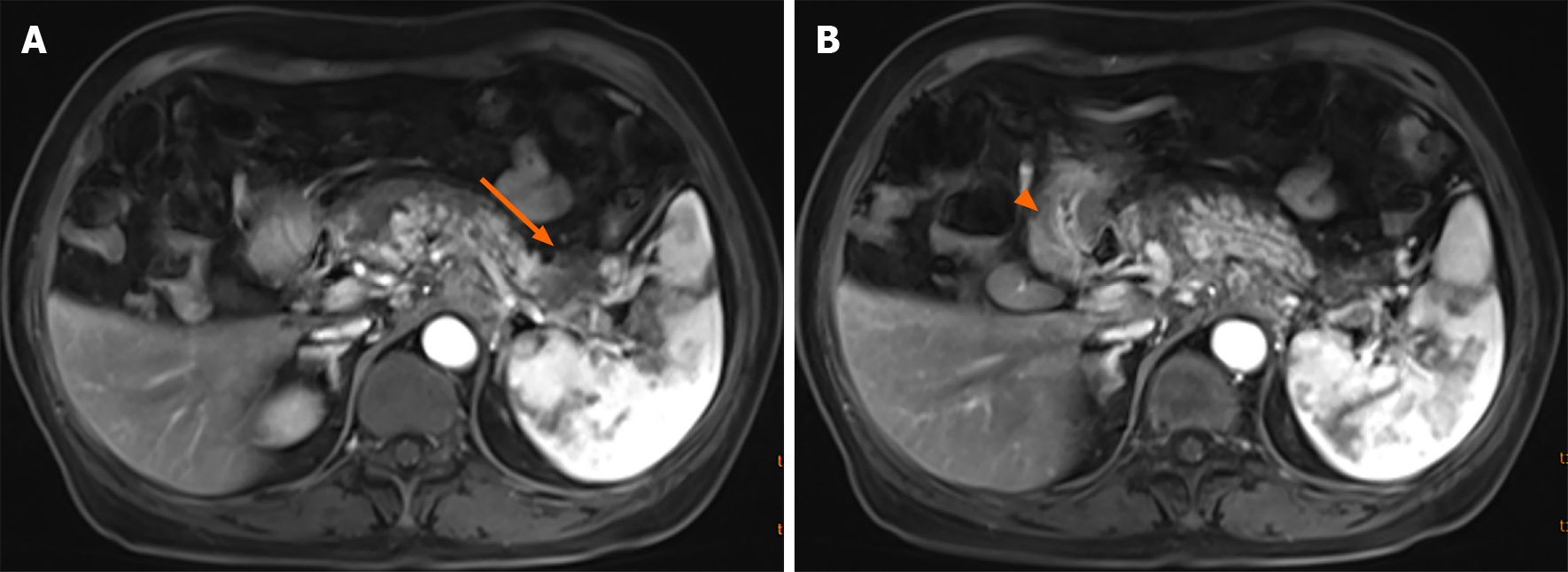
Figure 4 Enhanced upper abdominal magnetic resonance imaging.
A: An intensified mass (orange arrow); B: Thickening of the pyloric wall (orange arrowhead).
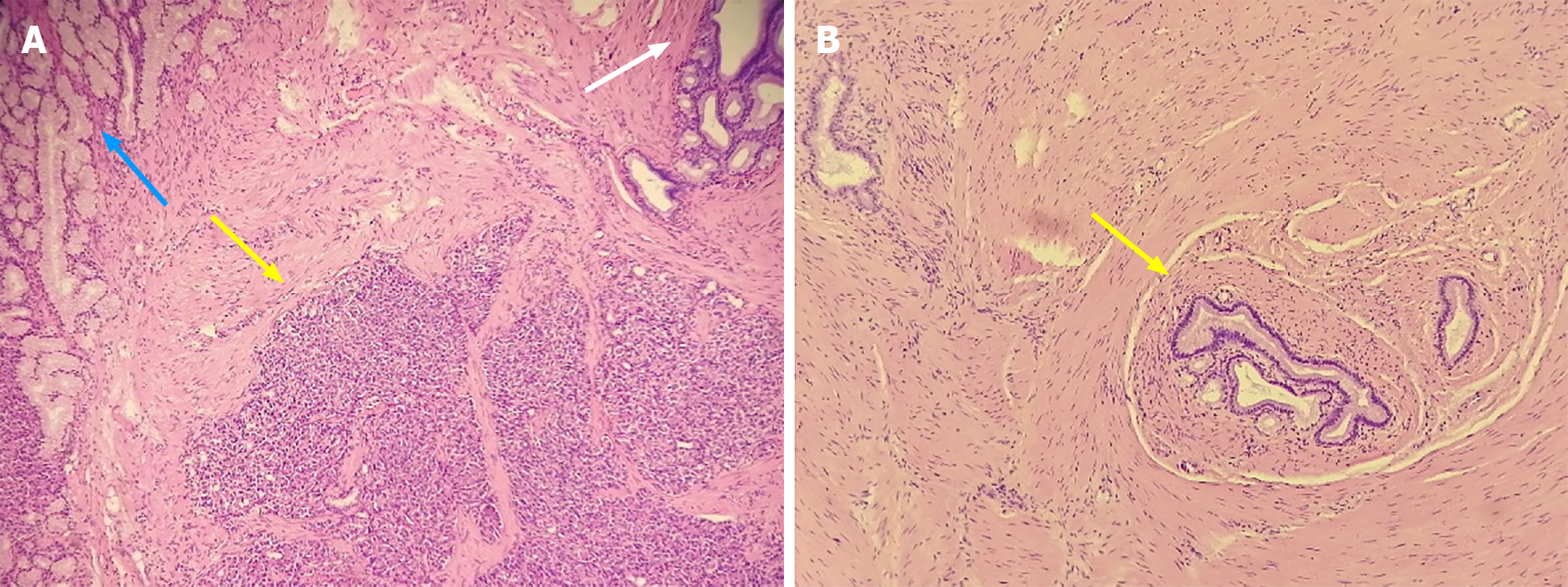
Figure 5 Histology (HE, 40 × magnification).
A: Disorganized pancreatic acini joining together and forming rough structures without islets and separated by bundles of smooth muscle (yellow arrow); the concomitant Brunner’s glands (blue arrow); and the undifferentiated mucus-secreting ducts, somewhat similar to gastric glands (white arrow); B: Typical gastric adenomyoma in another section of the gastric mass (yellow arrow) showing a mucus-secreting duct lined with columnar or cubic epithelial cells and surrounded by proliferating smooth muscle cells without heterotopic pancreas nearby.
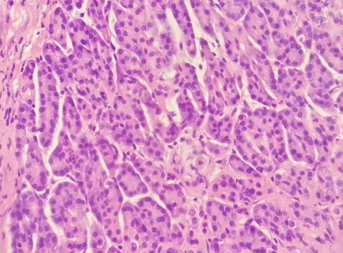
Figure 6 Histology (HE, 200 × magnification).
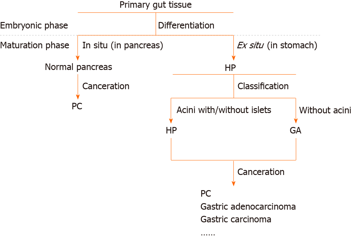
Figure 7 Flow chart of differentiation from primary gut tissue into normal pancreas, heterotopic pancreas, and gastric adenomyoma.
PC: Pancreatic cancer; HP: Heterotopic pancreas; GA: Gastric adenomyoma.
- Citation: Li K, Xu Y, Liu NB, Shi BM. Asymptomatic gastric adenomyoma and heterotopic pancreas in a patient with pancreatic cancer: A case report and review of the literature. World J Clin Cases 2021; 9(27): 8147-8156
- URL: https://www.wjgnet.com/2307-8960/full/v9/i27/8147.htm
- DOI: https://dx.doi.org/10.12998/wjcc.v9.i27.8147


