Copyright
©The Author(s) 2021.
World J Clin Cases. Sep 26, 2021; 9(27): 8120-8126
Published online Sep 26, 2021. doi: 10.12998/wjcc.v9.i27.8120
Published online Sep 26, 2021. doi: 10.12998/wjcc.v9.i27.8120
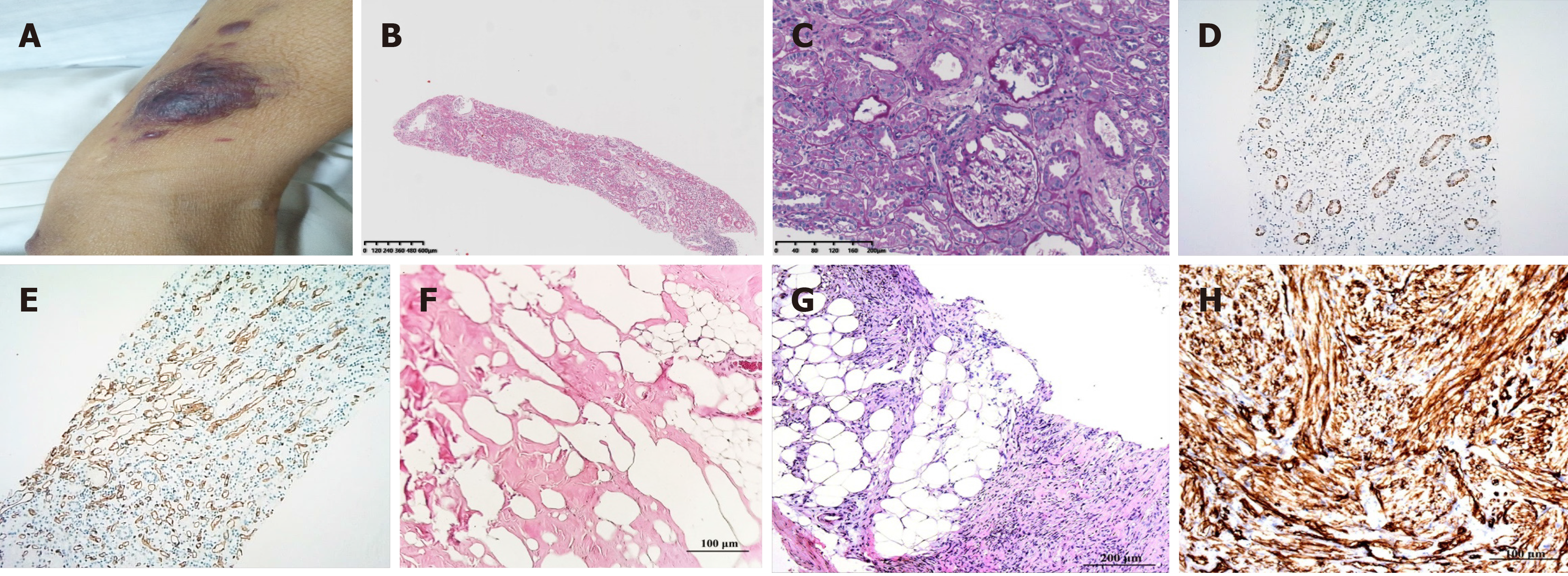
Figure 1 Histological features of the kidney biopsy and dermatological manifestation.
A: Infiltrative plaques are scattered throughout the body; B: Hematoxylin and eosin (H&E) staining of kidney biopsy; C: Periodic acid-silver methenamine staining of the kidney; D: Representative immunohistochemical staining for CD34 in the kidney; E: Representative immunohistochemical staining for CD117 in the kidney; F: H&E staining of the lymph node; G: H&E staining of the skin; H: Representative immunohistochemical staining for CD34.
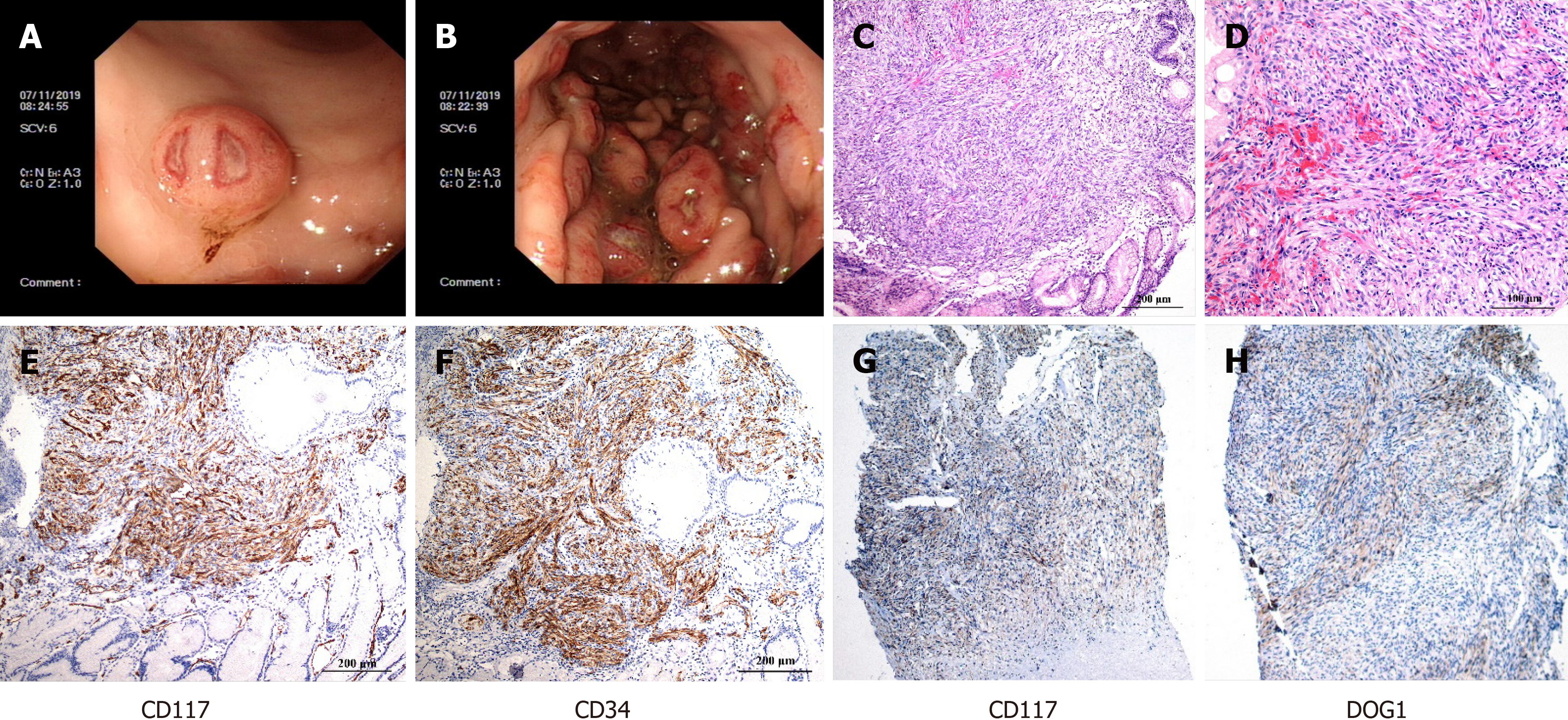
Figure 2 Gastroscopic and pathological findings of the tumors.
A: A representative photomicrograph was taken by gastroscopy, showing a mucosal mass with a diameter of 0.6 cm on the greater curvature of the stomach; B: A representative photomicrograph was taken by gastroscopy, showing 0.4-2 cm masses in the anterior wall of the middle and upper part of the gastric fundus; C: Hematoxylin and eosin (H&E) staining of the muscular layer of the gastric wall showing that the tumor was composed of spindle cells; D: H&E staining of epithelioid cells was occasionally seen with 6-8 mitotic figures/50 high power fields; E: Representative immunohistochemical staining for CD117 in the stomach; F: Representative immunohistochemical staining for CD34 in the stomach; G: Representative immunohistochemical staining for CD117 in the skin; H: Representative immunohistochemical staining for DOG1 in the skin.
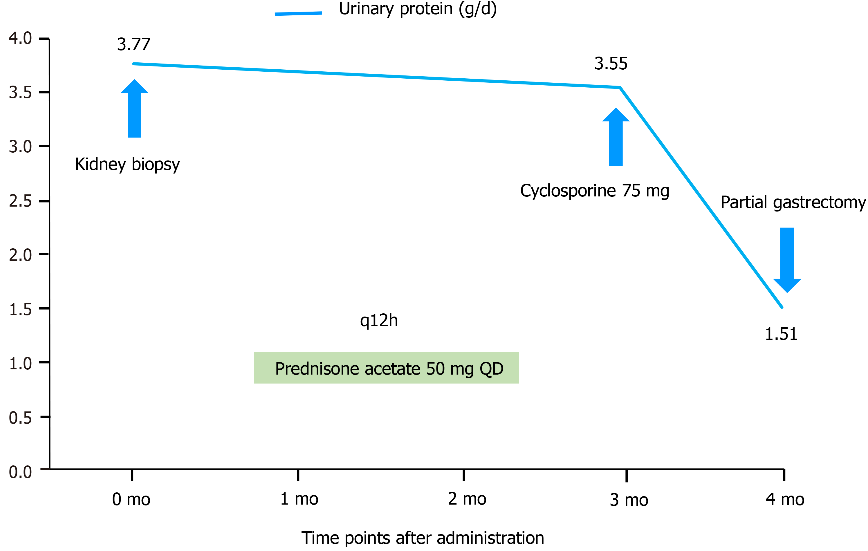
Figure 3 The patient’s clinical course.
A graph of the urine protein levels after the administration is shown.
- Citation: Zhou J, Yang Z, Yang CS, Lin H. Paraneoplastic focal segmental glomerulosclerosis associated with gastrointestinal stromal tumor with cutaneous metastasis: A case report. World J Clin Cases 2021; 9(27): 8120-8126
- URL: https://www.wjgnet.com/2307-8960/full/v9/i27/8120.htm
- DOI: https://dx.doi.org/10.12998/wjcc.v9.i27.8120


