Copyright
©The Author(s) 2021.
World J Clin Cases. Sep 26, 2021; 9(27): 8114-8119
Published online Sep 26, 2021. doi: 10.12998/wjcc.v9.i27.8114
Published online Sep 26, 2021. doi: 10.12998/wjcc.v9.i27.8114
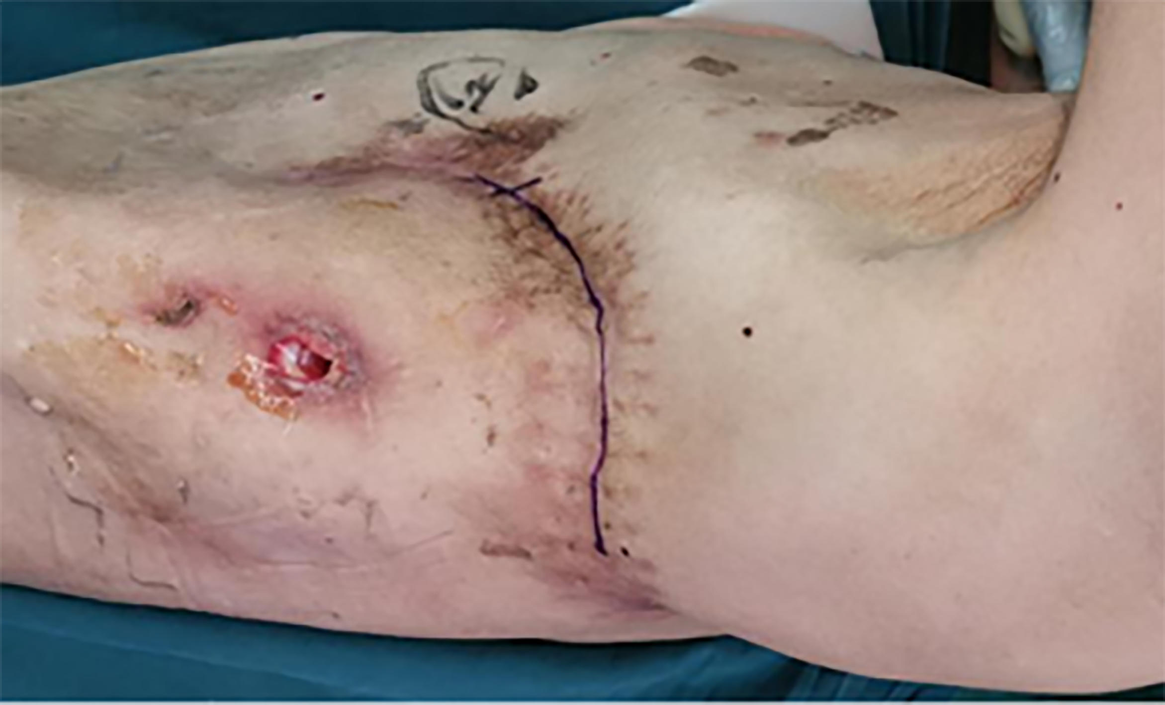
Figure 1 Preoperative image showing a posterolateral scarred incision on the left collapsed thorax.
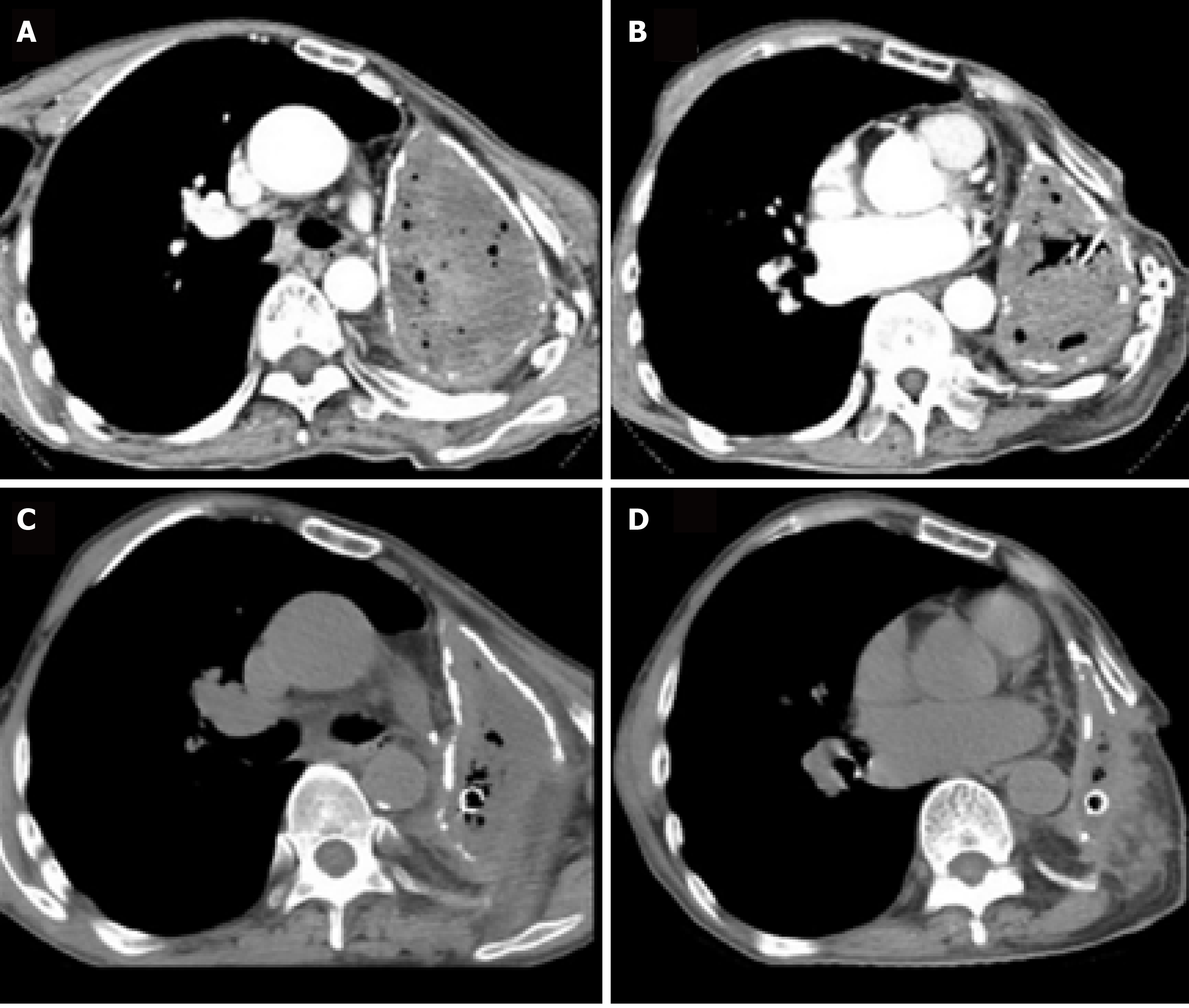
Figure 2 Chest computed tomography scan.
A and B: Preoperative chest computed tomography (CT) scan (axial view) revealed complete opacification of the left contracted chest; C and D: Postoperative chest CT scan (axial view) showed that the pleural volume was reduced by half at two months after limited thoracoplasty.
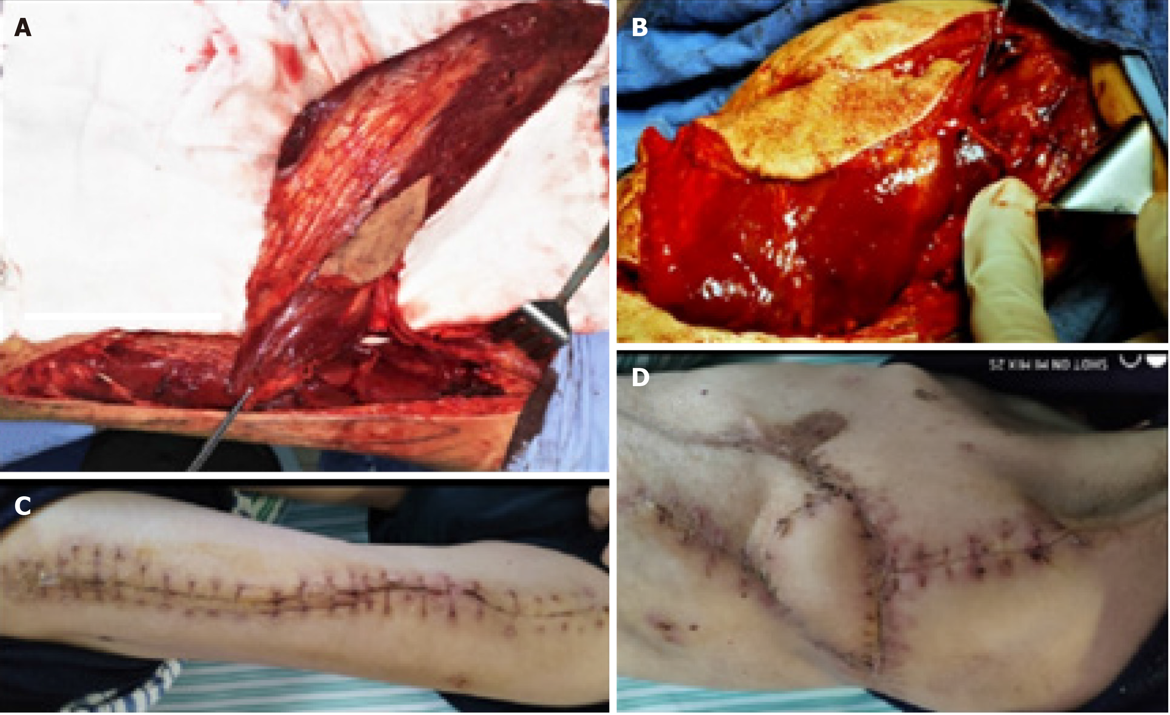
Figure 3 Intraoperative and postoperative images.
A: A 27 cm × 11 cm free vastus lateralis musculocutaneous flap was harvested from the patient’s left thigh; B: The free flap was transferred to obliterate the residual pleural space; C: Healed donor site; D: The free musculocutaneous flap healed very well.
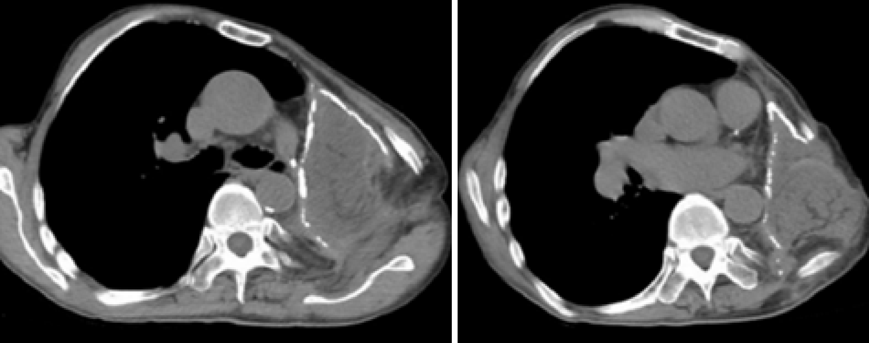
Figure 4 Postoperative chest computed tomography scan (axial view) showing successful obliteration of the empyema cavity by limited thoracoplasty and free vastus lateralis musculocutaneous flap transposition.
- Citation: Huang QQ, He ZL, Wu YY, Liu ZJ. Limited thoracoplasty and free musculocutaneous flap transposition for postpneumonectomy empyema: A case report. World J Clin Cases 2021; 9(27): 8114-8119
- URL: https://www.wjgnet.com/2307-8960/full/v9/i27/8114.htm
- DOI: https://dx.doi.org/10.12998/wjcc.v9.i27.8114


