Copyright
©The Author(s) 2021.
World J Clin Cases. Sep 26, 2021; 9(27): 8097-8103
Published online Sep 26, 2021. doi: 10.12998/wjcc.v9.i27.8097
Published online Sep 26, 2021. doi: 10.12998/wjcc.v9.i27.8097
Figure 1 Preoperative axial views of high-resolution computed tomography venography of the diploic vein.
A: The cephalic diploic vein (orange arrow) runs in the parietal bone; B: The middle portion (orange arrow) runs in the parietal bone; C: The caudal portion (orange arrow) passes through the mastoid air cells posteriorly in a dehiscent canal (blue arrows); D: The caudal portion (orange arrow) courses into the transverse-sigmoid sinus.
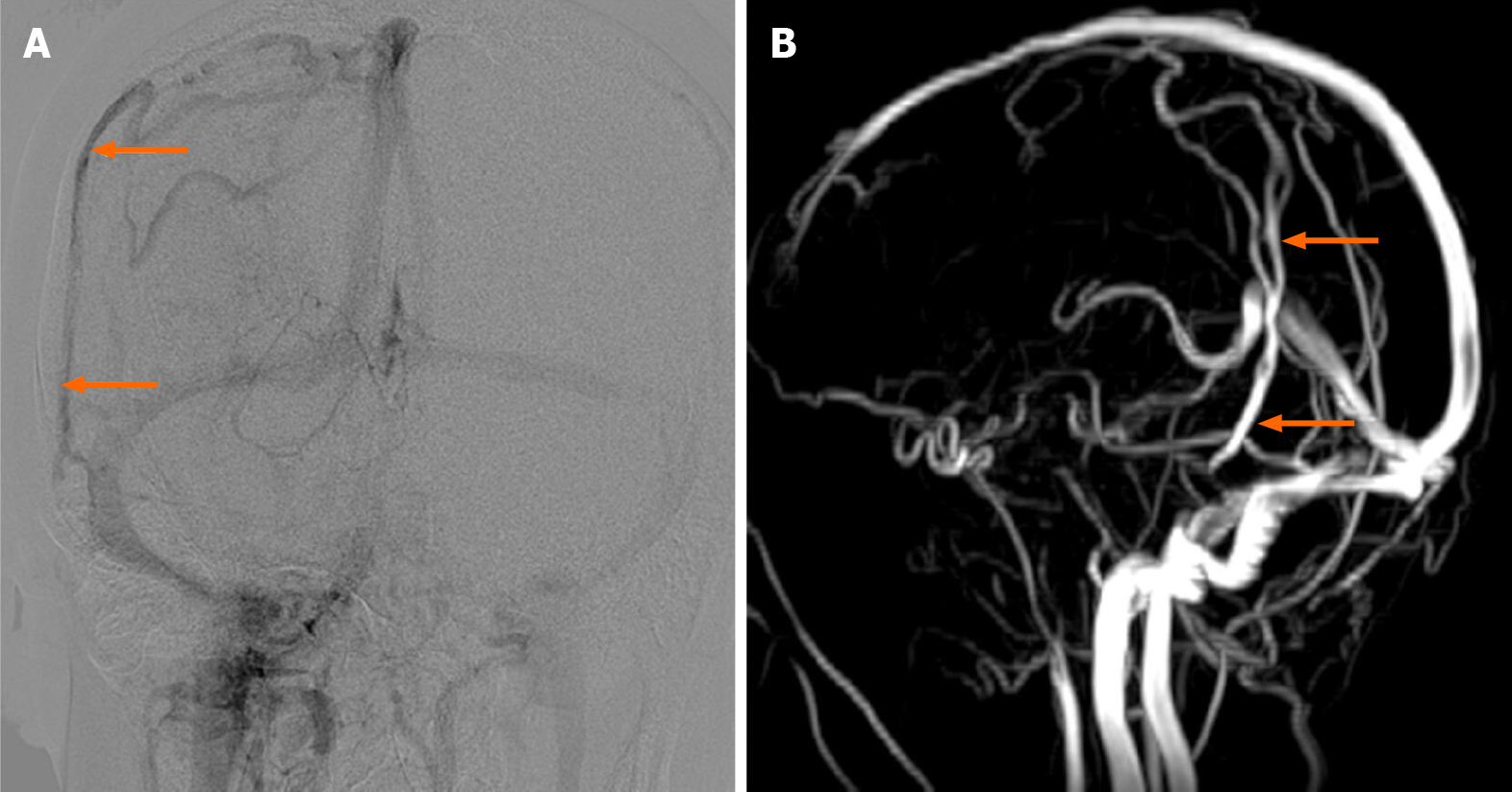
Figure 2 Preoperative images of the diploic vein.
A: Anteroposterior image of digital subtraction angiography; B: Lateral image of magnetic resonance venography.
Figure 3 Preoperative four-dimensional flow magnetic resonance view of the caudal diploic vein and adjacent transverse sinus and sigmoid sinus.
DV: Diploic vein; SS: Sigmoid sinus; TS: Transverse sinus; A: Anterior; P: Posterior; S: Superior; I: Inferior.
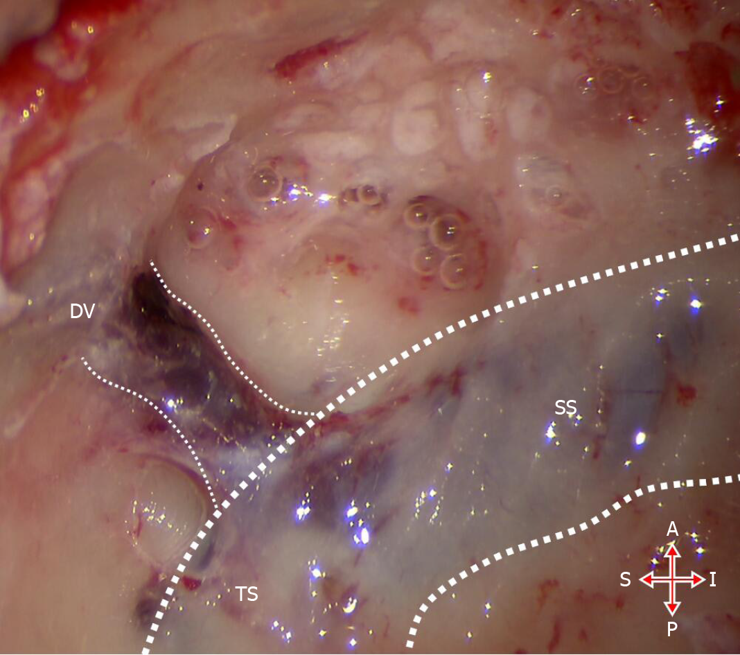
Figure 4 Intraoperative view.
DV: Diploic vein; SS: Sigmoid sinus; TS: Transverse sinus; A: Anterior; P: Posterior; S: Superior; I: Inferior.
- Citation: Zhao PF, Zeng R, Qiu XY, Ding HY, Lv H, Li XS, Wang GP, Li D, Gong SS, Wang ZC. Diploic vein as a newly treatable cause of pulsatile tinnitus: A case report. World J Clin Cases 2021; 9(27): 8097-8103
- URL: https://www.wjgnet.com/2307-8960/full/v9/i27/8097.htm
- DOI: https://dx.doi.org/10.12998/wjcc.v9.i27.8097


