Copyright
©The Author(s) 2021.
World J Clin Cases. Sep 26, 2021; 9(27): 8082-8089
Published online Sep 26, 2021. doi: 10.12998/wjcc.v9.i27.8082
Published online Sep 26, 2021. doi: 10.12998/wjcc.v9.i27.8082
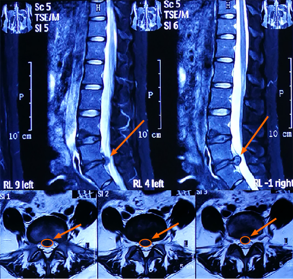
Figure 1 Magnetic resonance images obtained upon admission.
The orange arrows indicate the location of disc herniation.
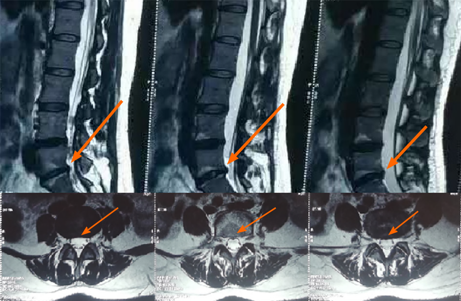
Figure 2 Magnetic resonance images obtained at 15 mo after discharge from hospital.
The orange arrows show the location of disc herniation retraction.
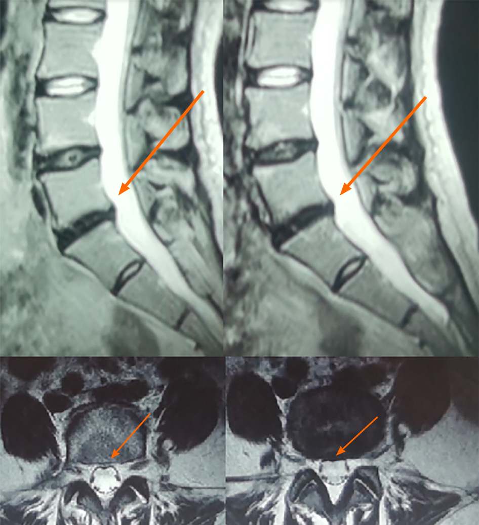
Figure 3 Magnetic resonance images obtained at 44 mo after discharge from hospital.
The orange arrows show the location of disc herniation retraction.
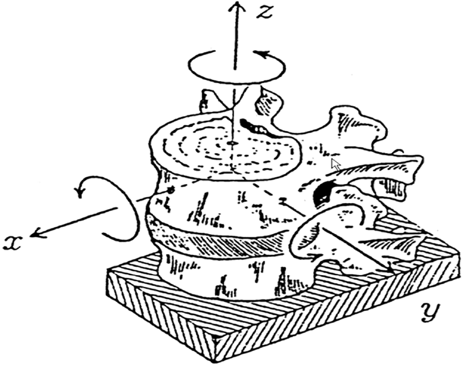
Figure 4
Direction of spinal movement.
- Citation: Wang P, Chen C, Zhang QH, Sun GD, Wang CA, Li W. Retraction of lumbar disc herniation achieved by noninvasive techniques: A case report. World J Clin Cases 2021; 9(27): 8082-8089
- URL: https://www.wjgnet.com/2307-8960/full/v9/i27/8082.htm
- DOI: https://dx.doi.org/10.12998/wjcc.v9.i27.8082


