Copyright
©The Author(s) 2021.
World J Clin Cases. Sep 16, 2021; 9(26): 7937-7943
Published online Sep 16, 2021. doi: 10.12998/wjcc.v9.i26.7937
Published online Sep 16, 2021. doi: 10.12998/wjcc.v9.i26.7937
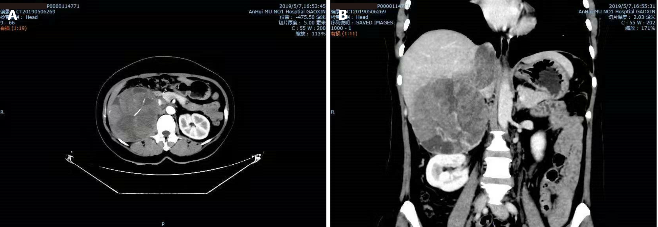
Figure 1 Computed tomography images showing a huge mass in the right adrenal gland in both transverse and coronal view.
A: Transverse view; B: Coronal view.
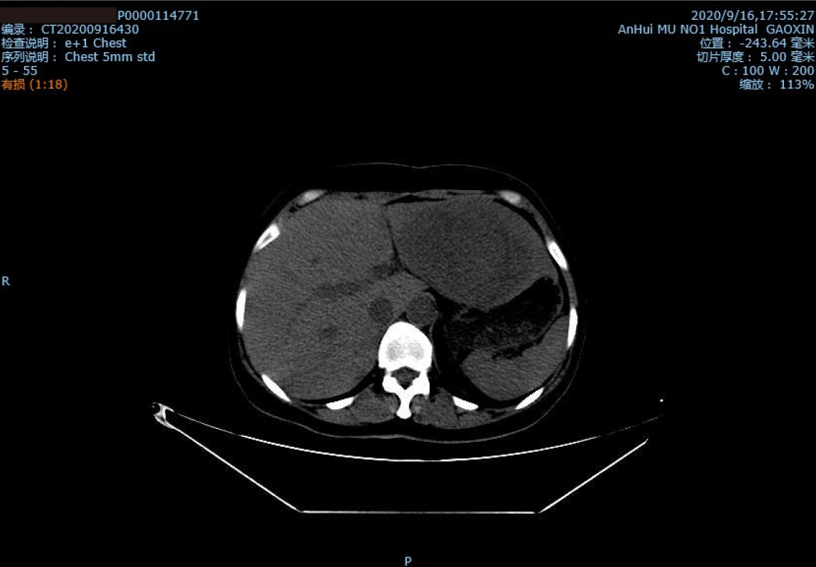
Figure 2 Computed tomography image showing a large, round, compact, and irregular mass on the left lobe of the liver, approximately 112.
7 mm × 79.8 mm.
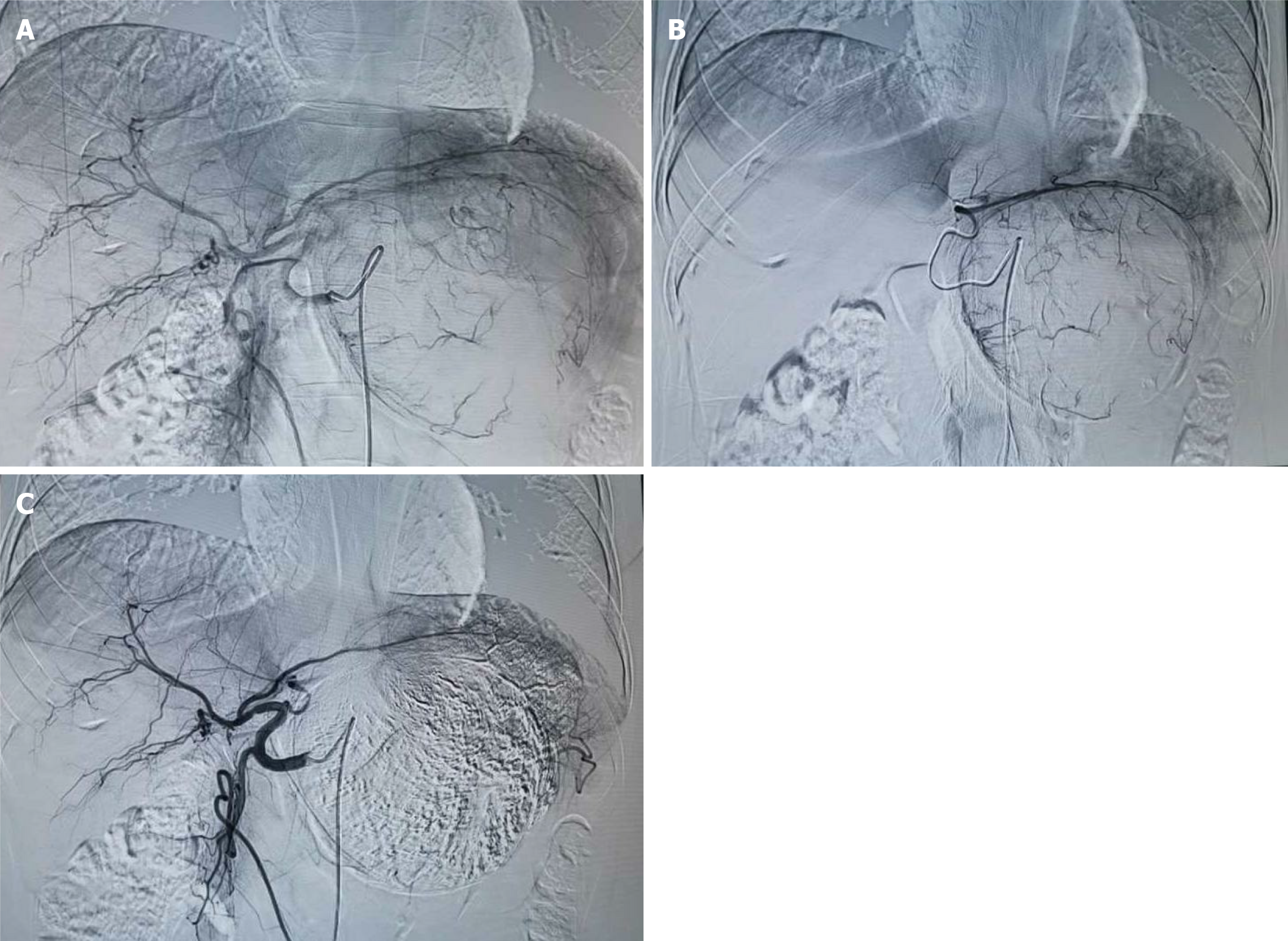
Figure 3 Intraoperative photographs.
A: There is a large, lightly stained tumor in the left lobe of the liver. The left hepatic artery is the main blood supply for the tumor; B: The guidewire entering the left hepatic artery for chemoembolization; C: The left lobe of the liver has a good deposit of lipiodol after the chemoembolization.
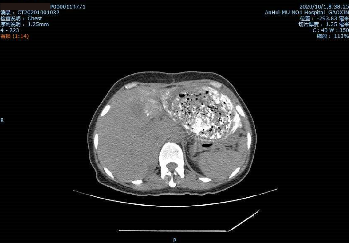
Figure 4 Computed tomography image showing a good uptake rate of lipiodol and honeycomb necrosis of liver tumor cells.
- Citation: Wang ZL, Sun X, Zhang FL, Wang T, Li P. Rare complication of acute adrenocortical dysfunction in adrenocortical carcinoma after transcatheter arterial chemoembolization: A case report. World J Clin Cases 2021; 9(26): 7937-7943
- URL: https://www.wjgnet.com/2307-8960/full/v9/i26/7937.htm
- DOI: https://dx.doi.org/10.12998/wjcc.v9.i26.7937


