Copyright
©The Author(s) 2021.
World J Clin Cases. Sep 16, 2021; 9(26): 7909-7916
Published online Sep 16, 2021. doi: 10.12998/wjcc.v9.i26.7909
Published online Sep 16, 2021. doi: 10.12998/wjcc.v9.i26.7909
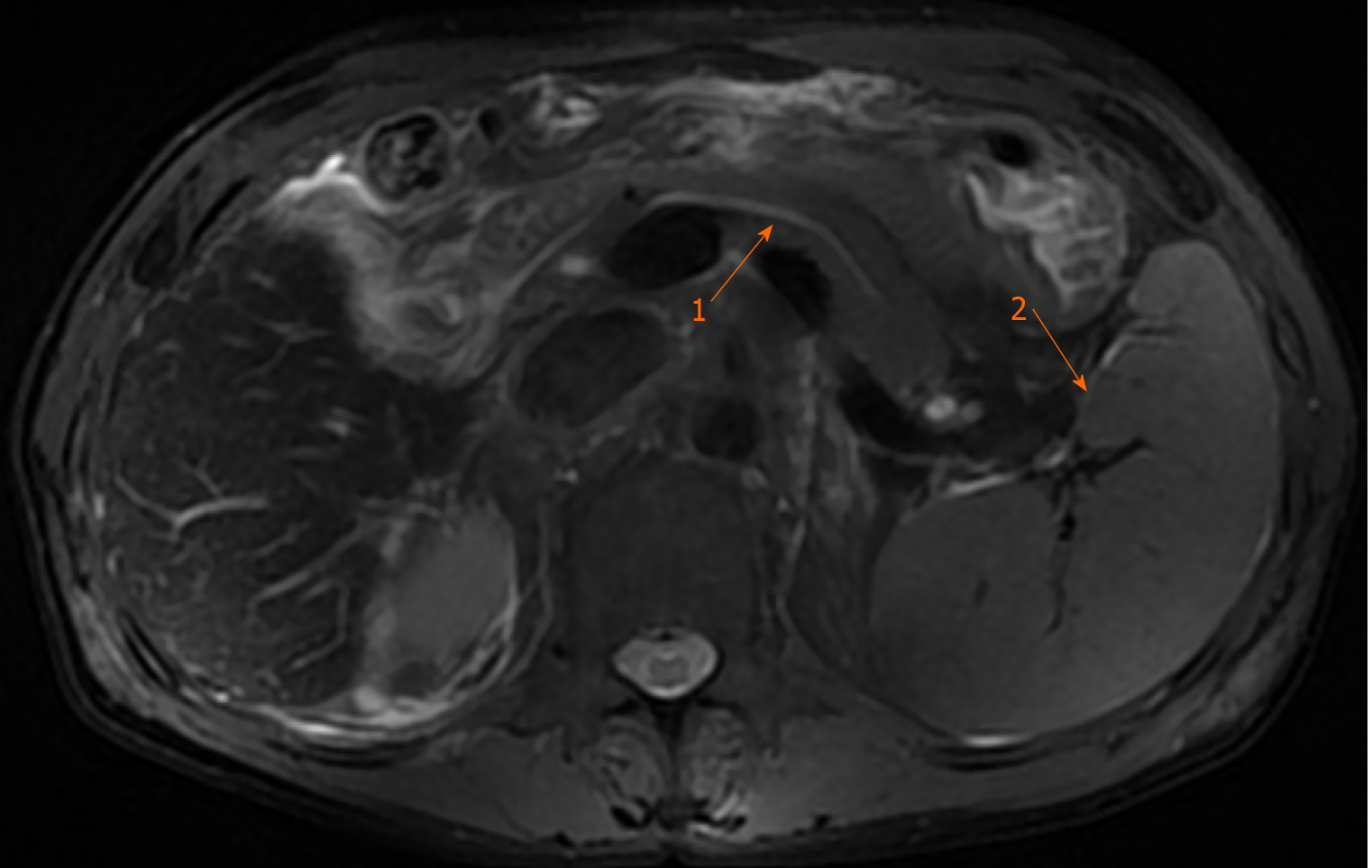
Figure 1 Magnetic resonance cholangiopancreatography of the upper abdomen revealed splenomegaly (arrow 2) and pancreatic duct dilatation (arrow 1).
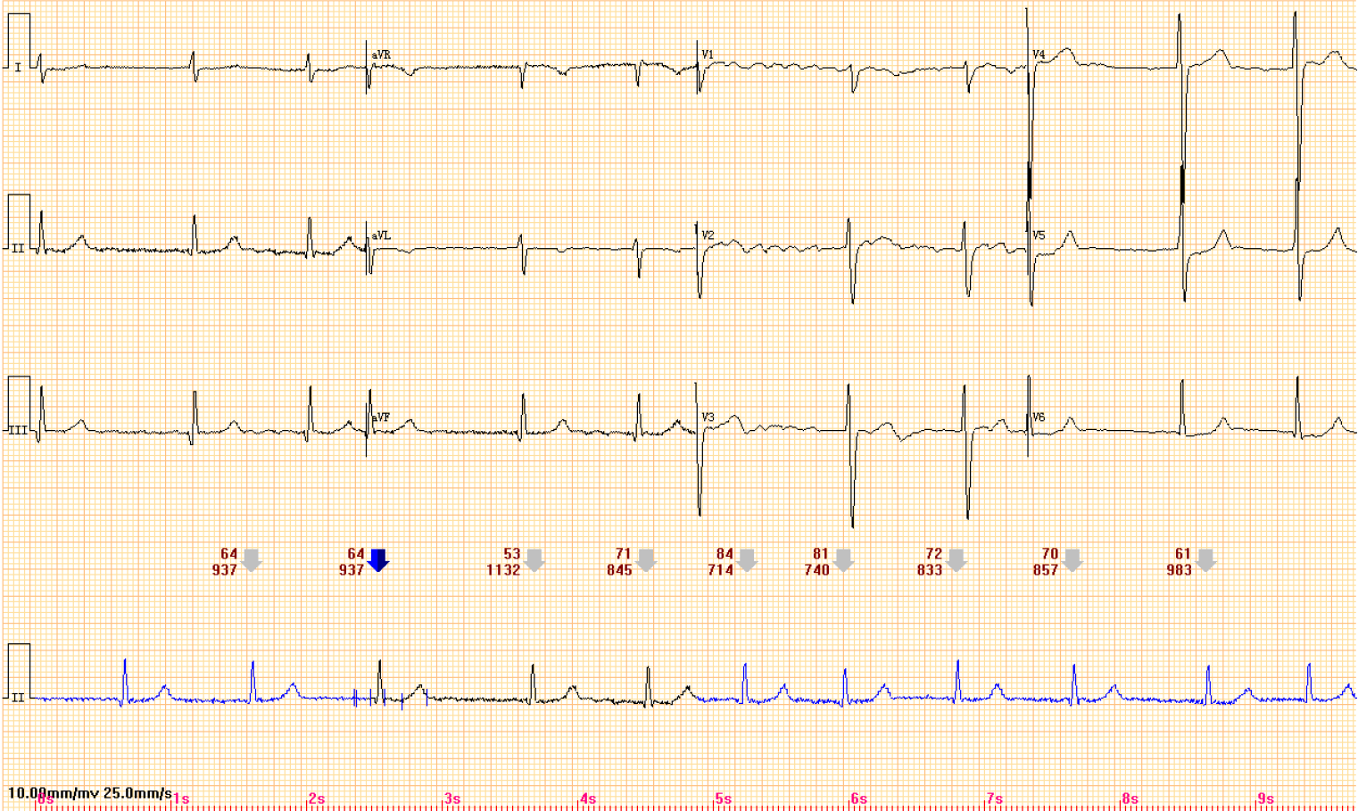
Figure 2 Electrocardiogram showing atrial fibrillation.
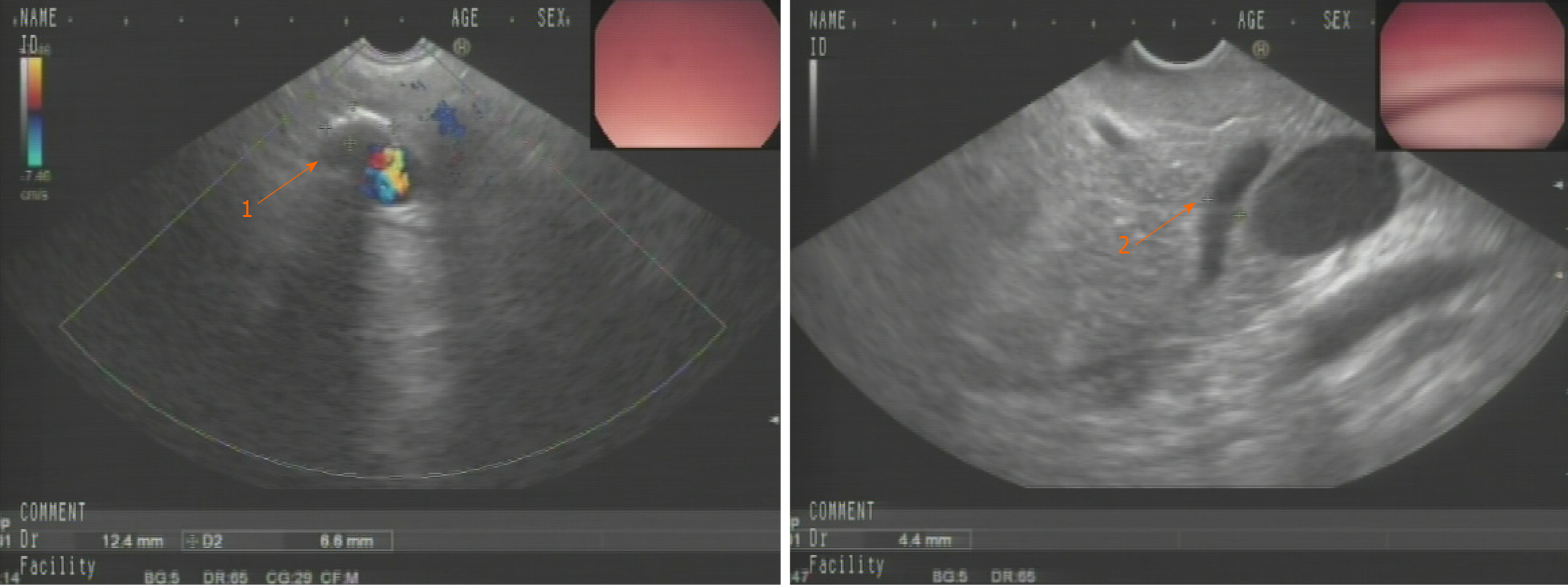
Figure 3 Ultrasound gastroscopy suggested the possibility of pancreatic duct stones (arrow 1) with dilatation of the pancreatic duct (arrow 2).
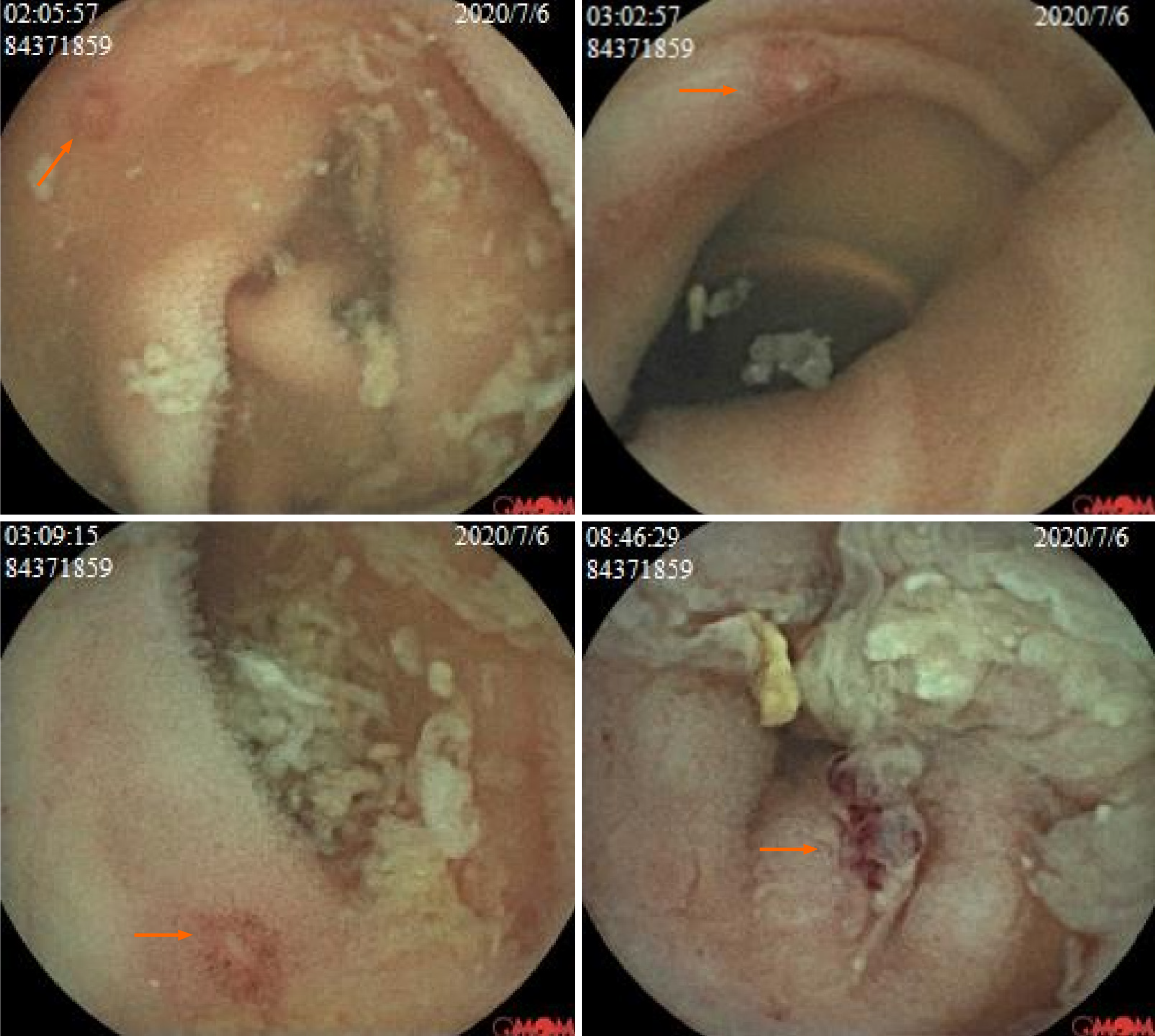
Figure 4 Capsule endoscopy revealed multiple intestinal ulcers (arrow).
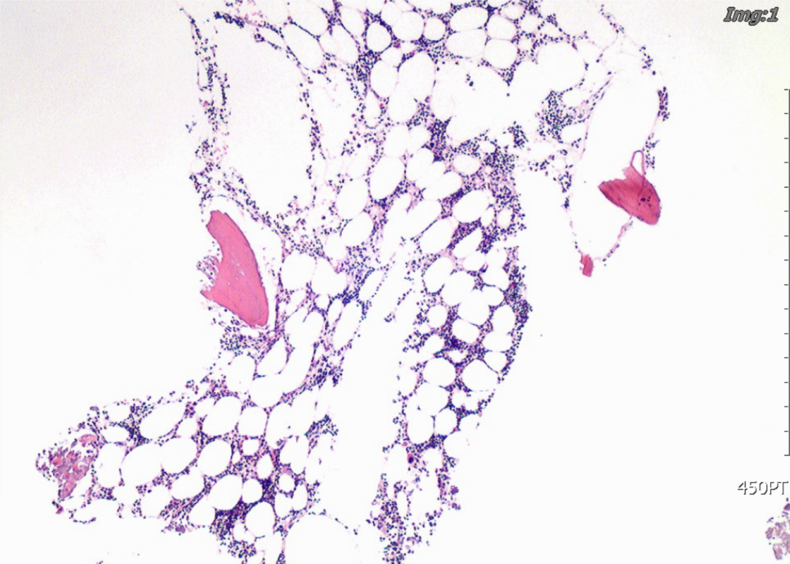
Figure 5 Bone marrow puncture smear of the iliac spine suggestive of hyperplastic anemia.
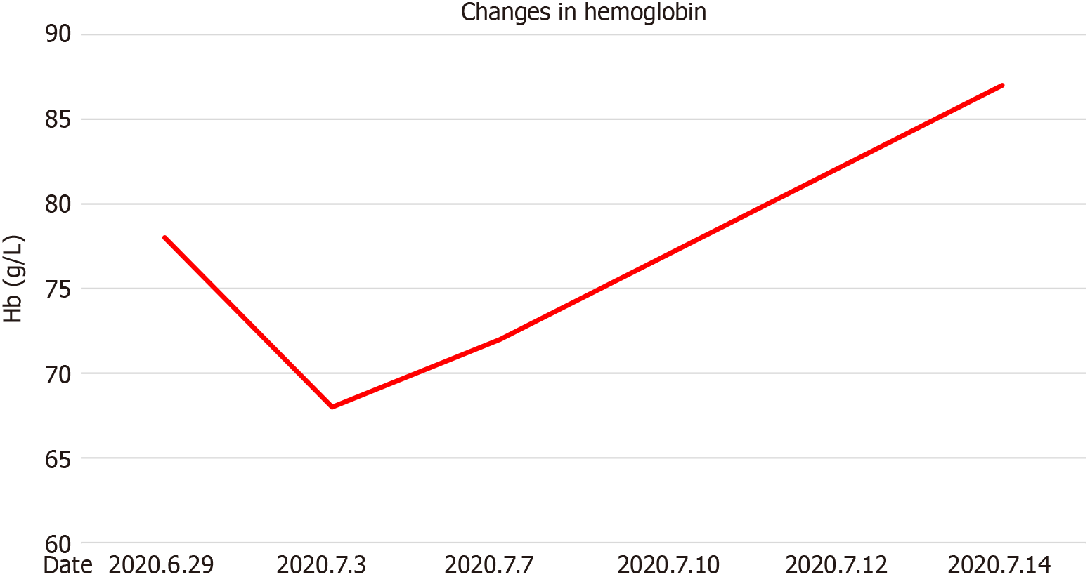
Figure 6 Changes in hemoglobin during hospitalization.
- Citation: Sun DJ, Li HT, Ye Z, Xu BB, Li DZ, Wang W. Gastrointestinal bleeding caused by syphilis: A case report. World J Clin Cases 2021; 9(26): 7909-7916
- URL: https://www.wjgnet.com/2307-8960/full/v9/i26/7909.htm
- DOI: https://dx.doi.org/10.12998/wjcc.v9.i26.7909


