Copyright
©The Author(s) 2021.
World J Clin Cases. Sep 16, 2021; 9(26): 7893-7900
Published online Sep 16, 2021. doi: 10.12998/wjcc.v9.i26.7893
Published online Sep 16, 2021. doi: 10.12998/wjcc.v9.i26.7893
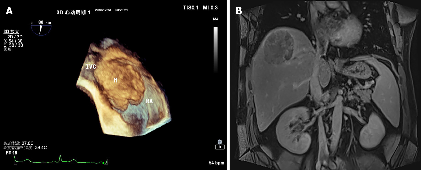
Figure 1 Preoperative imaging examination.
A: A medium echo was seen in the right atrium, with a clear boundary, local calcification on the surface, and wide base; B: Magnetic resonance imaging showed that there were filling defects in the hepatic vein and right atrium. IVC: Inferior vena cava; RA: Right atrium.
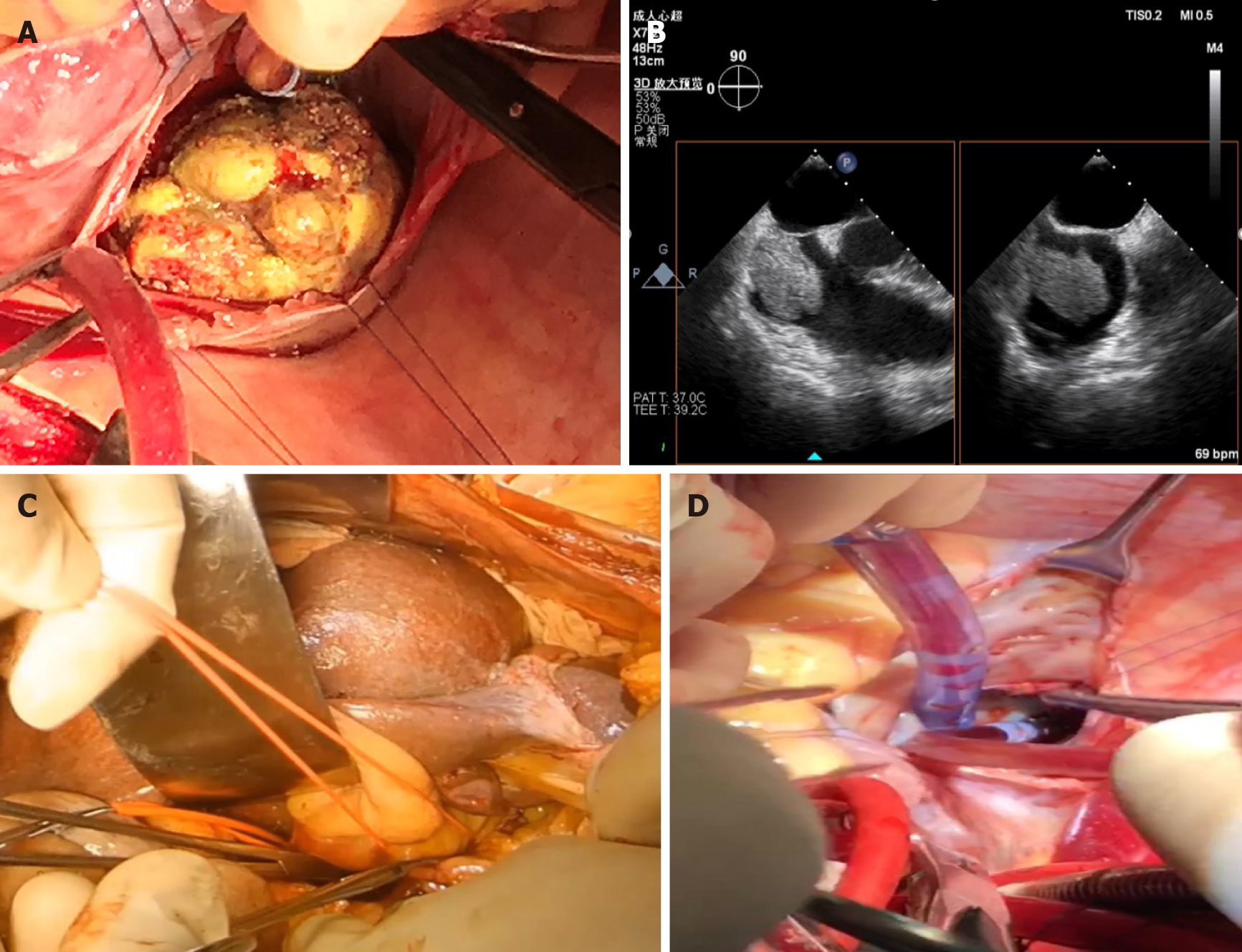
Figure 2 Images of surgical operation.
A: Image of head of the tumor thrombus; B: Monitored image of transesophageal echocardiography during the operation; C: The hepatic vascular exclusion was blocked for 16 min; D: The entrance of the inferior vena cava and the right atrium was clear.
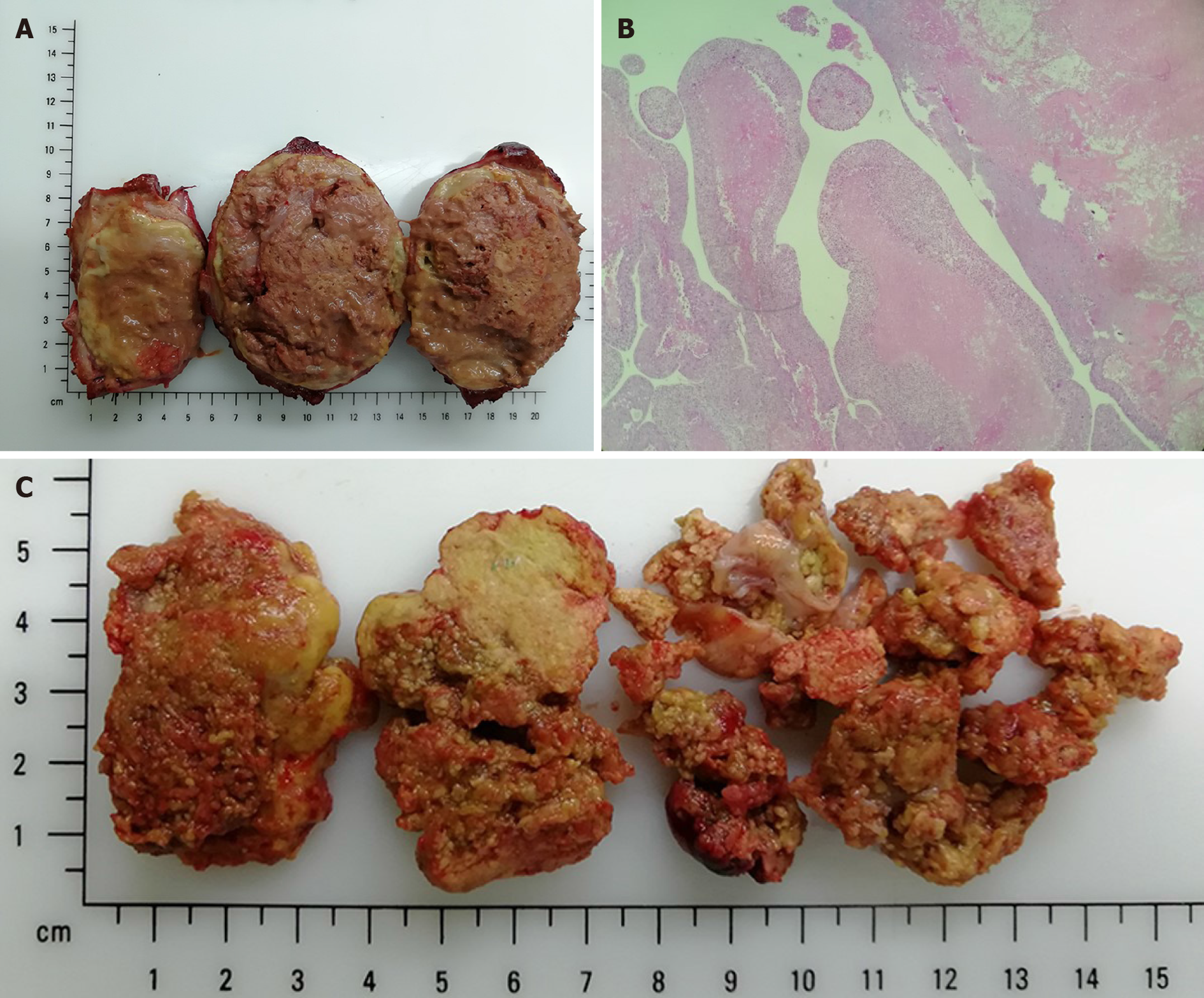
Figure 3 The shape and pathology of the tumor thrombus.
A: The tumor is about 10 cm × 8.5 cm × 8 cm, and most of the mass is necrotic; B: Active cancer cells could be seen as finger-like bulge around the focal capsule, and necrosis area was over 75% (magnification 200 ×); C: The atrium thrombus is 15 cm × 5 cm × 3 cm with a long, thin neck.
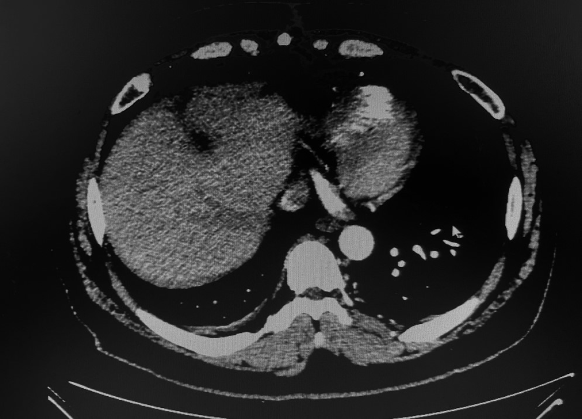
Figure 4
There was no tumor recurrence or metastasis over a 2-year follow-up.
- Citation: Liu J, Zhang RX, Dong B, Guo K, Gao ZM, Wang LM. Hepatocellular carcinoma with inferior vena cava and right atrium thrombus: A case report. World J Clin Cases 2021; 9(26): 7893-7900
- URL: https://www.wjgnet.com/2307-8960/full/v9/i26/7893.htm
- DOI: https://dx.doi.org/10.12998/wjcc.v9.i26.7893


