Copyright
©The Author(s) 2021.
World J Clin Cases. Sep 16, 2021; 9(26): 7886-7892
Published online Sep 16, 2021. doi: 10.12998/wjcc.v9.i26.7886
Published online Sep 16, 2021. doi: 10.12998/wjcc.v9.i26.7886
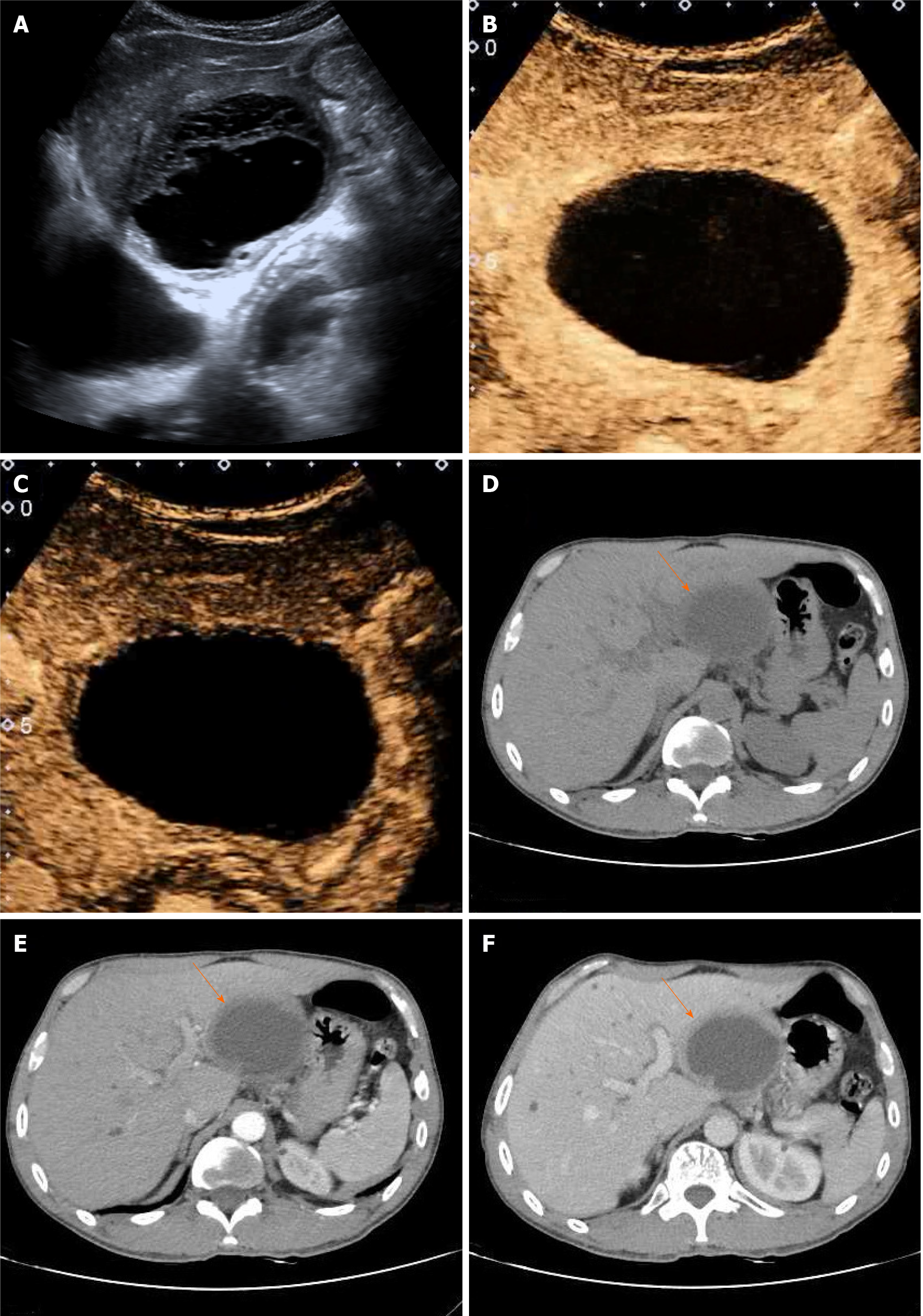
Figure 1 Ultrasound and computerized tomography imaging of intrahepatic biliary cystadenoma.
A: Ultrasound (US) showed that the thick-walled anechoic area in the left lateral lobe was regular and had a clear boundary; B: Arterial phase contrast-enhanced US (CEUS) showed that the anechoic area presented peripheral annular hyperenhancement and that the central part and internal septations showed no enhancement; C: Portal phase CEUS showed that the enhanced cyst wall subsided slowly but was still above the surrounding liver tissue; D: Computerized tomography revealed a round thick-walled monolocular mass (arrow) with clear borders and uniform density of cyst fluid but no visible separation in the left lateral lobe; E: In the arterial phase, the cyst wall was enhanced, but the cyst fluid was not enhanced (arrow); F: In the portal phase, homogeneous enhancement was observed for the cyst wall close to the surrounding normally enhanced liver tissue (arrow).
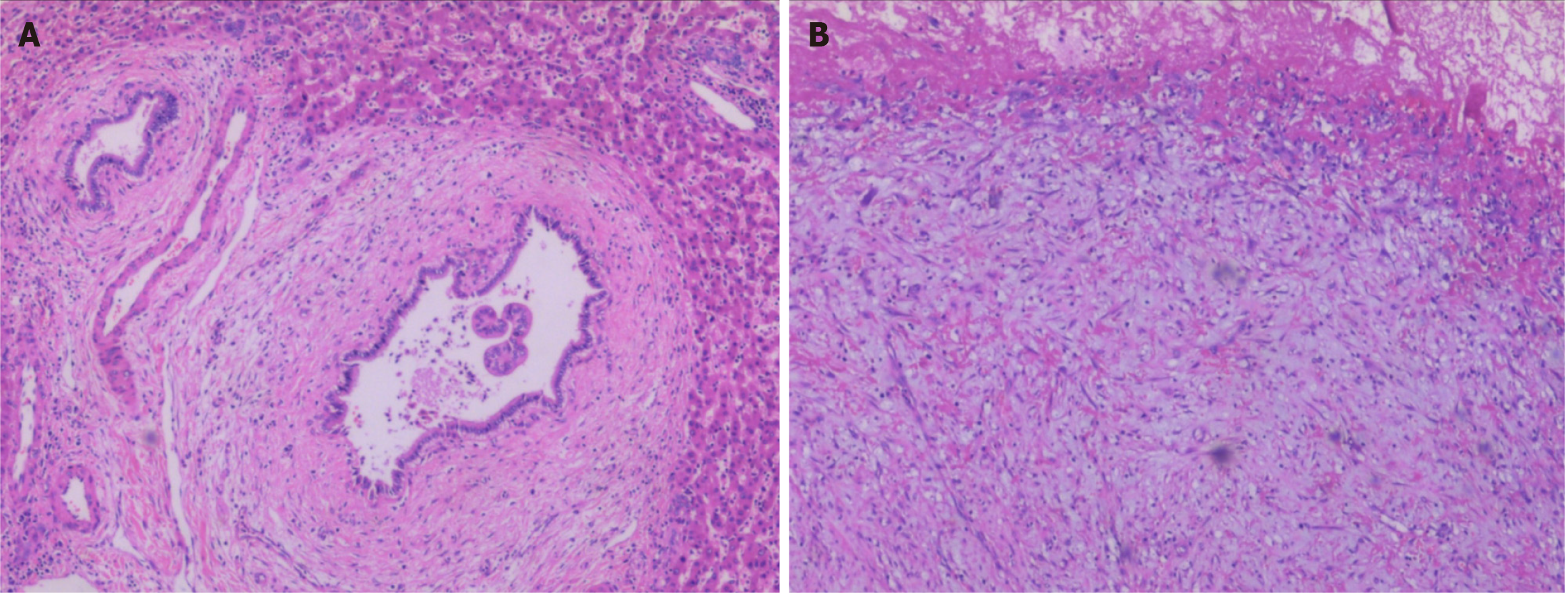
Figure 2 Postoperative pathological image of Intrahepatic biliary cystadenoma.
A: The tumor wall was lined with a single layer of cuboidal or columnar epithelial cells that were arranged regularly without conspicuous atypia. Fibrinoid necrosis was observed in the capsule (hematoxylin and eosin stain, × 200); B: Fibroplasia and mucinous degeneration presented in the subepithelial stroma, along with acute and chronic inflammatory cell infiltration (hematoxylin and eosin stain, × 200).
- Citation: Che CH, Zhao ZH, Song HM, Zheng YY. Rare monolocular intrahepatic biliary cystadenoma: A case report. World J Clin Cases 2021; 9(26): 7886-7892
- URL: https://www.wjgnet.com/2307-8960/full/v9/i26/7886.htm
- DOI: https://dx.doi.org/10.12998/wjcc.v9.i26.7886


