Copyright
©The Author(s) 2021.
World J Clin Cases. Sep 16, 2021; 9(26): 7876-7885
Published online Sep 16, 2021. doi: 10.12998/wjcc.v9.i26.7876
Published online Sep 16, 2021. doi: 10.12998/wjcc.v9.i26.7876
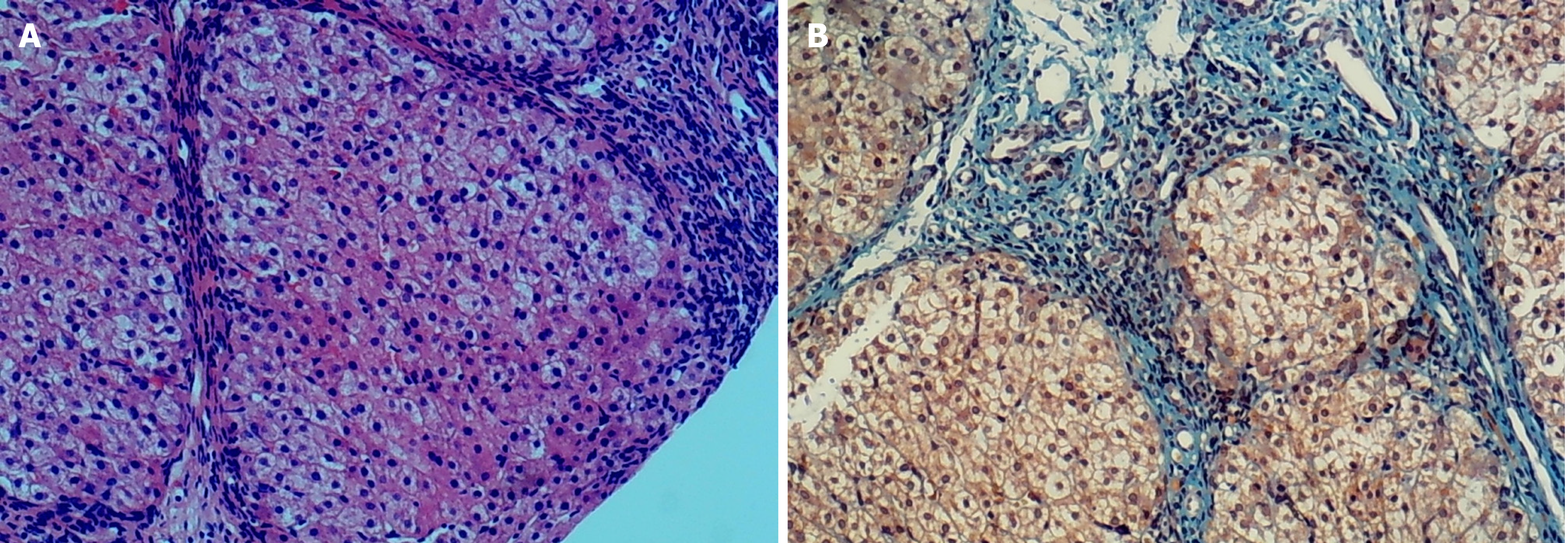
Figure 1 Liver tissue hematoxylin and eosin staining and Masson staining.
A: Hematoxylin and eosin staining of liver tissue suggested the presence of pseudo lobules. Original magnification (100 ×); B: Collagen fiber staining (Masson) of liver tissue in green suggested fibrous hyperplasia. Original magnification (100 ×).
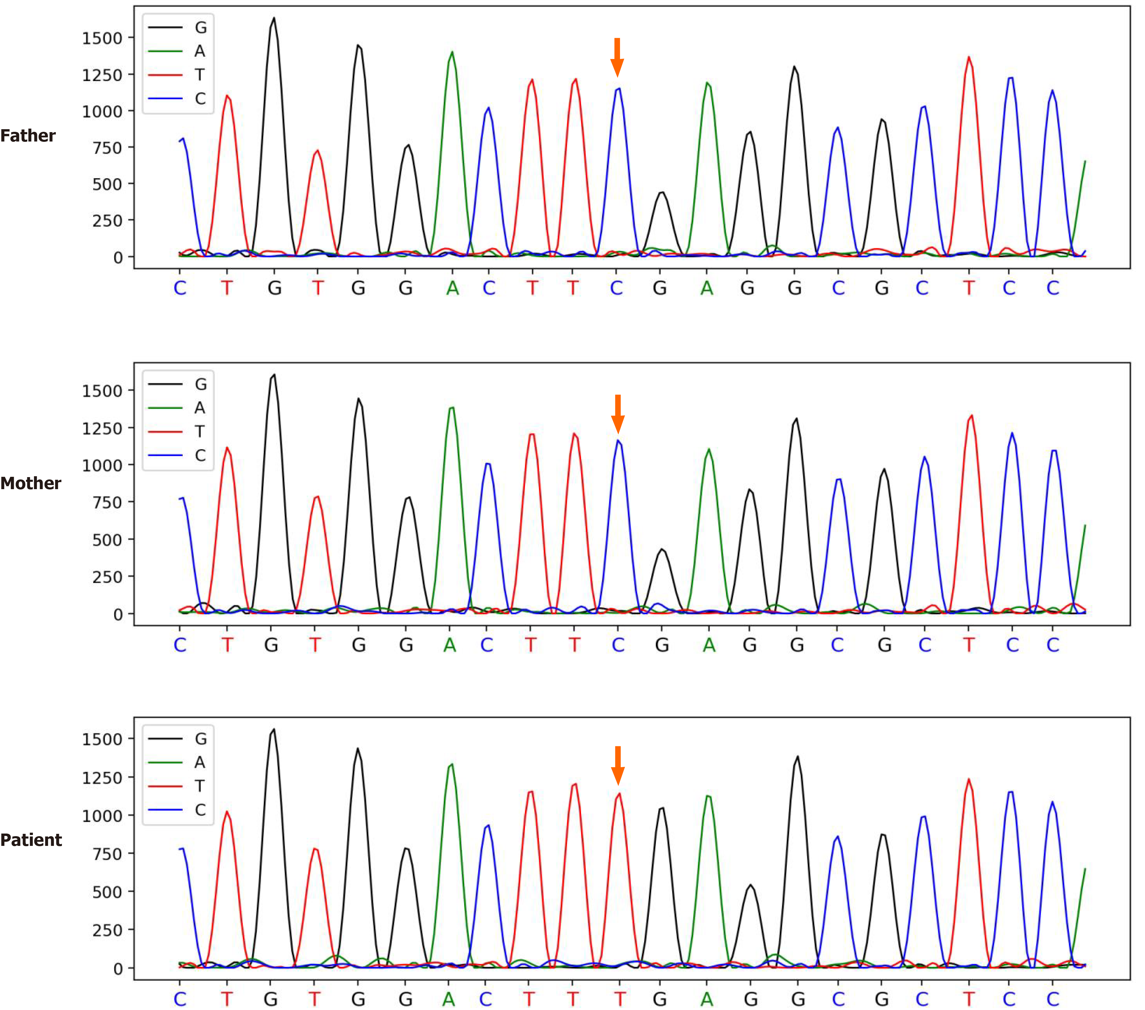
Figure 2 Genetic testing results of the family.
The patient carries the c.1036G>A (p.E346K) mutation in the ATP6AP1 gene. The red arrow refers to mutated nucleotide c.1036G>A in the patient. The genotypes of this locus in his parents were normal.
- Citation: Yang X, Lv ZL, Tang Q, Chen XQ, Huang L, Yang MX, Lan LC, Shan QW. Congenital disorder of glycosylation caused by mutation of ATP6AP1 gene (c.1036G>A) in a Chinese infant: A case report. World J Clin Cases 2021; 9(26): 7876-7885
- URL: https://www.wjgnet.com/2307-8960/full/v9/i26/7876.htm
- DOI: https://dx.doi.org/10.12998/wjcc.v9.i26.7876


