Copyright
©The Author(s) 2021.
World J Clin Cases. Sep 16, 2021; 9(26): 7863-7869
Published online Sep 16, 2021. doi: 10.12998/wjcc.v9.i26.7863
Published online Sep 16, 2021. doi: 10.12998/wjcc.v9.i26.7863
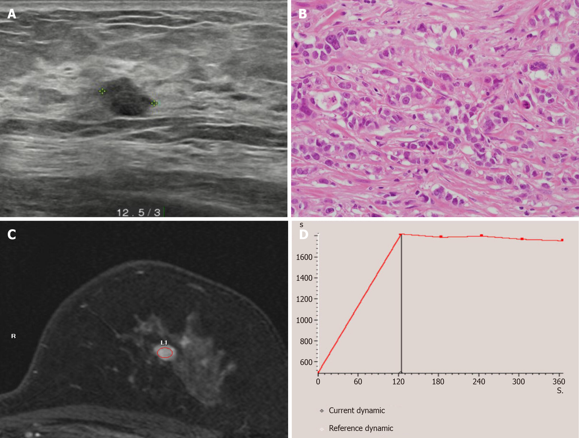
Figure 1 Radiologic findings of the left breast mass diagnosed as invasive ductal carcinoma.
A: Breast ultrasonography showed an 0.8 cm × 0.7 cm sized irregular hypoechoic mass located on the left 12:30 o’clock position at 3 cm distance from the left nipple; B: Breast core needle biopsy showed invasive ductal carcinoma with no special type (Haematoxylin and eosin staining × 400); C and D: Breast magnetic resonance imaging showed single enhancing mass on the left breast mass with a type II dynamic curve.
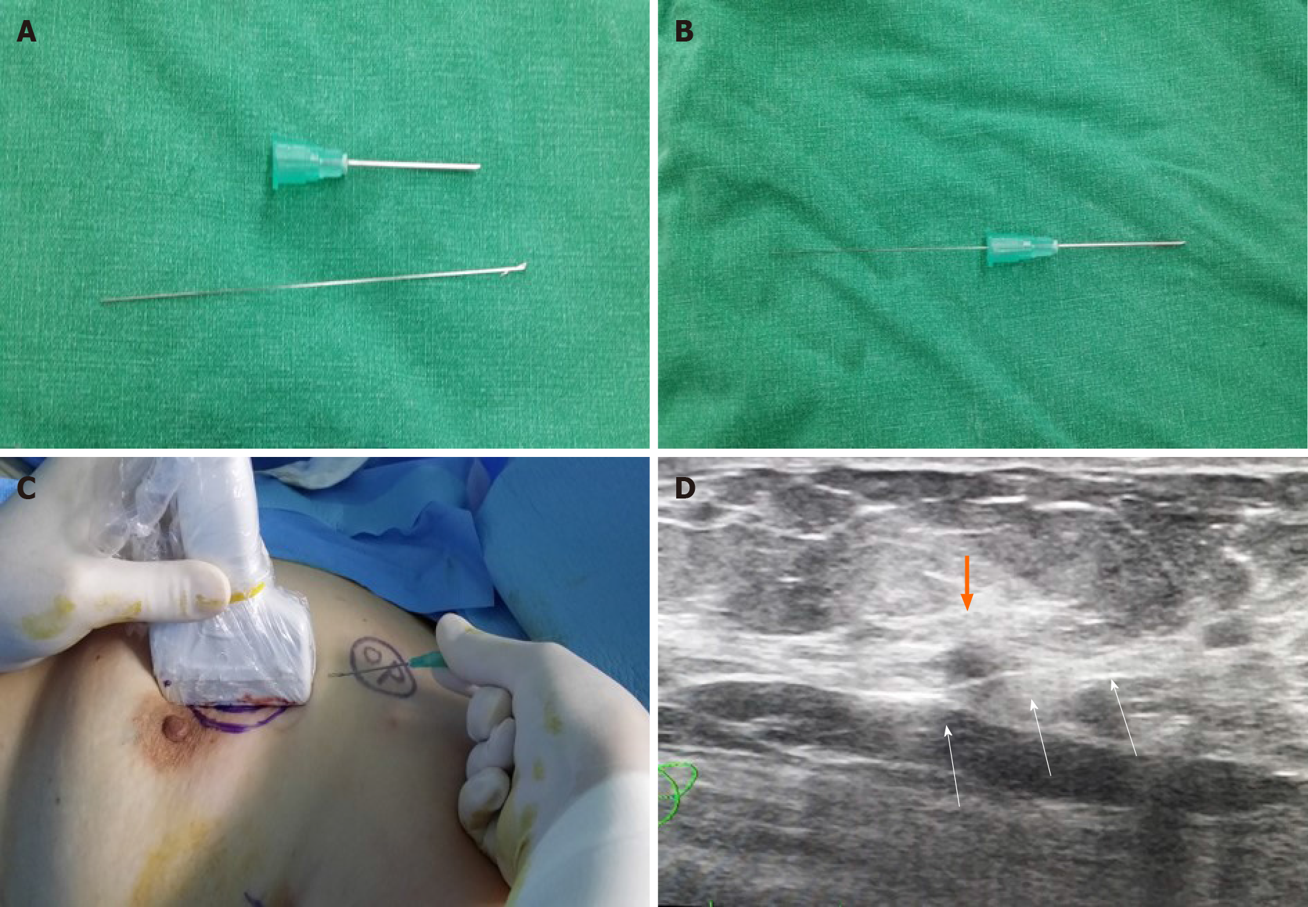
Figure 2 Method of intraoperative ultrasound-guided wire localization before operation.
A and B: The breast lesion localization wire consisted of a 23-gauze needle, through which a 25-gauze, 10 cm long monofilament wire with a distal hook; C and D: The wire was inserted and left in the breast as the needle was totally withdrawn. The three white arrows indicates the localized wire (white arrows) that was inserted to left breast mass (orange).
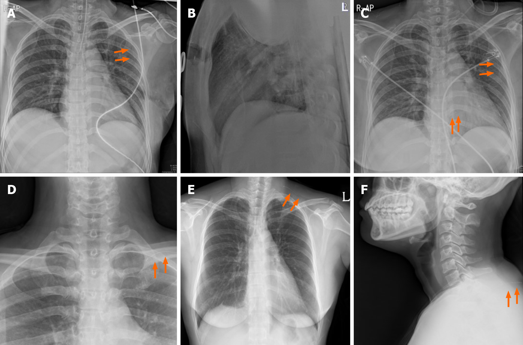
Figure 3 Serial simple X-ray image of the patient.
A and B: Intraoperative portable chest posteroanterior X-ray revealed the wire (orange arrows) to be located on midaxillary line-level longitudinally. But, it was not detected on lateral film; C: Recovery room portable chest posteroanterior X-ray revealed that the wire was located on midaxillary line-level longitudinally. This was the same finding in the intraoperative chest posteroanterior X-ray. No pneumothorax was seen; D: One day after the operation, a neck X-ray revealed the wire was located on the level of the clavicle; E and F: Two days after the operation, a serial simple X-ray revealed that the wire was located on a subcutaneous lesion of the back.
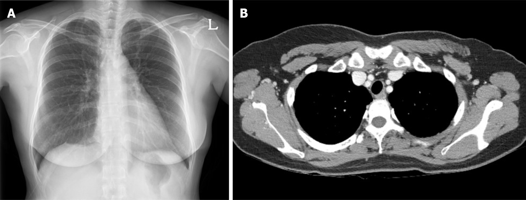
Figure 4 Follow up images of the patient.
A: Three months after surgery, a chest posteroanterior X-ray; B: Chest computed tomography revealed no evidence of remnant wire or pneumothorax and other abnormalities.
- Citation: Choi YJ. Migration of the localization wire to the back in patient with nonpalpable breast carcinoma: A case report. World J Clin Cases 2021; 9(26): 7863-7869
- URL: https://www.wjgnet.com/2307-8960/full/v9/i26/7863.htm
- DOI: https://dx.doi.org/10.12998/wjcc.v9.i26.7863


