Copyright
©The Author(s) 2021.
World J Clin Cases. Sep 16, 2021; 9(26): 7798-7804
Published online Sep 16, 2021. doi: 10.12998/wjcc.v9.i26.7798
Published online Sep 16, 2021. doi: 10.12998/wjcc.v9.i26.7798
Figure 1 Gastroscopy before treatment, gastric retention (black arrows) can be seen.
Figure 2 Hematoxylin and eosin staining showed macrophage (black arrows) infiltration in the lamina propria.
A: Original magnification 200 ×; B: Original magnification 400 ×.
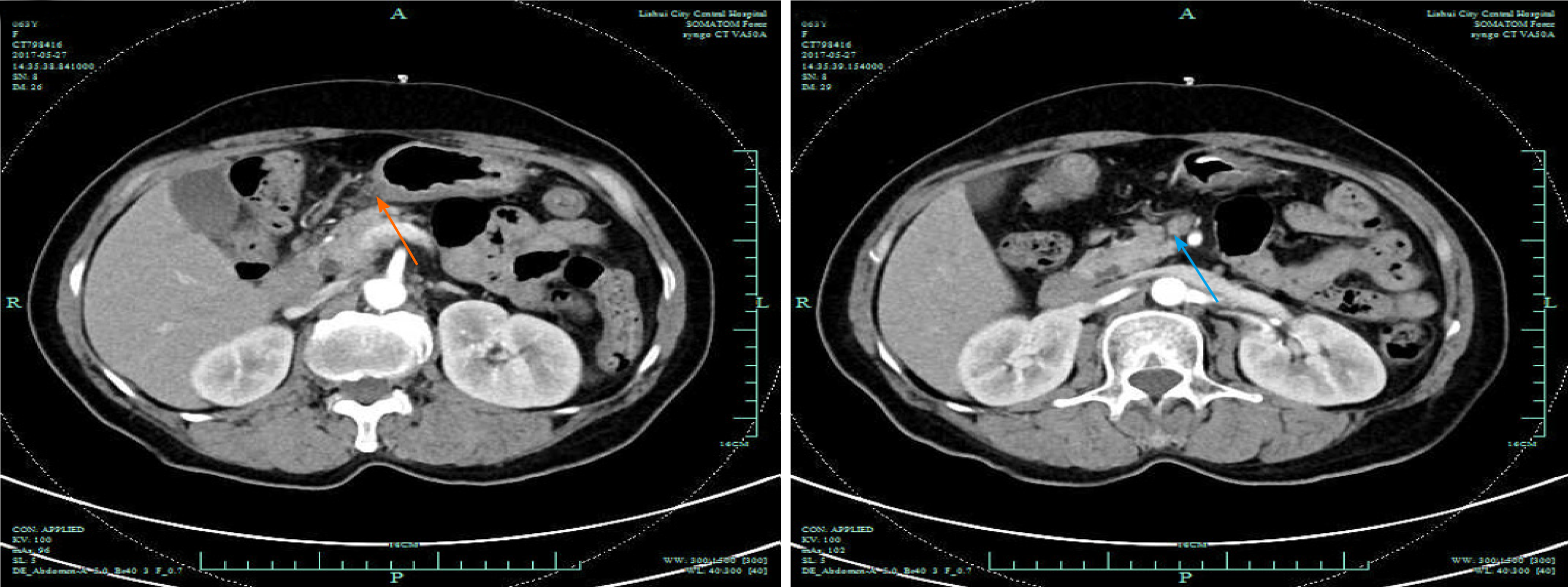
Figure 3 The gastric wall of the antrum was thickened (orange arrow), and some enlarged lymph nodes (blue arrow) were seen in the abdominal cavity, retroperitoneum, and bilateral groin areas.
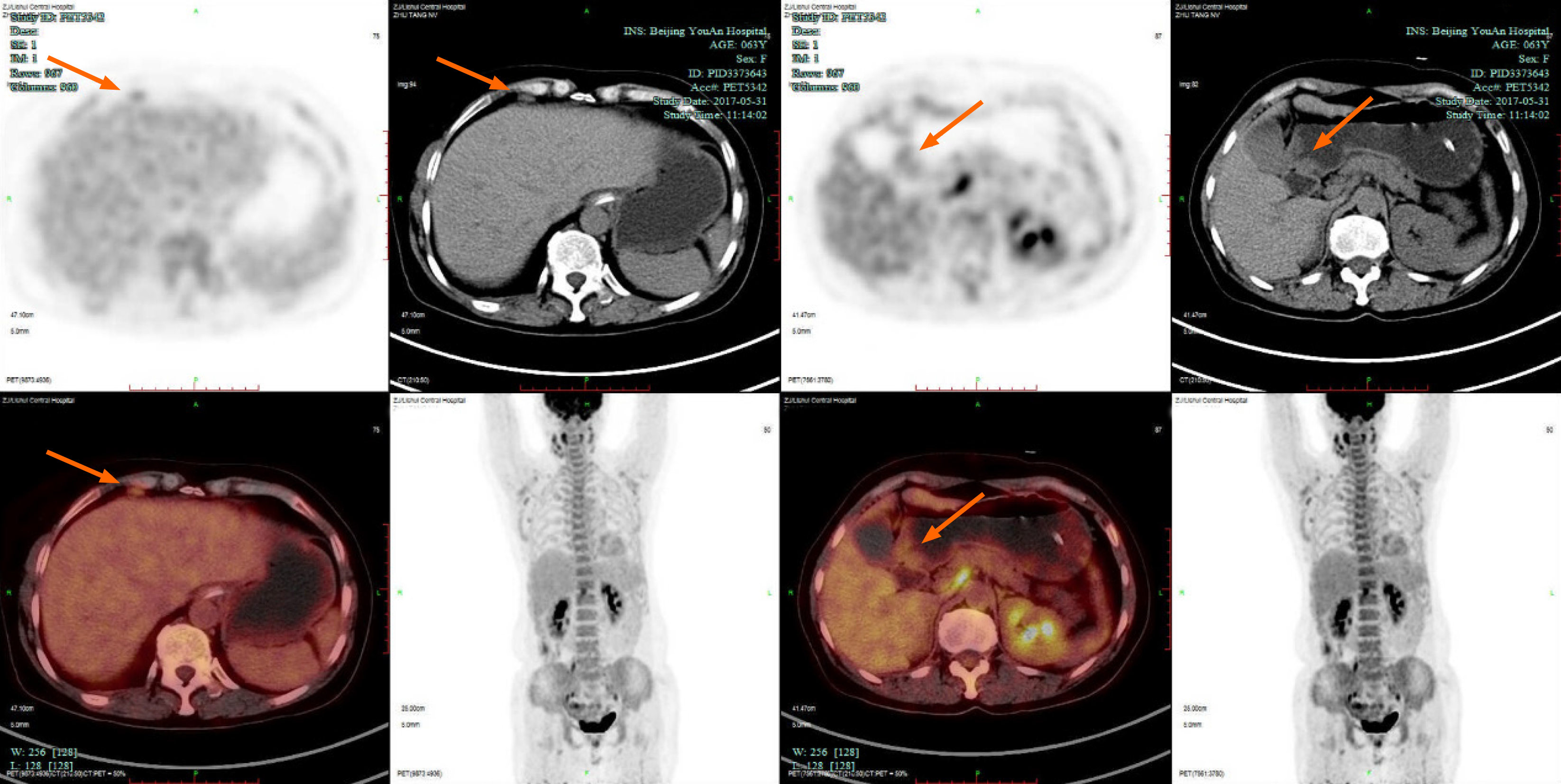
Figure 4 Positron emission tomography/computed tomography: The walls of the gastric antrum and pylorus were locally thickened, and the tracer uptake was increased.
The maximum standardized uptake value was 2.4. Multiple lymphadenopathies (orange arrows) can be seen in the perigastric and left supraclavicular areas, right anterior diaphragm, right retroperitoneum, iliac vessels and pelvic area.
Figure 5 Endoscopic ultrasound revealed an antral gastric ulcer, with a low-echo thickened mucosa (green arrows), measuring 8.
1 mm at its thickest. The muscularis propria was conserved.
Figure 6 A second gastroscopy showed antral ulcer (black arrows) in the lesser curvature of gastric antrum.
Figure 7 Gastroscopy 2 mo after treatment.
Resolution of clinical symptoms and endoscopic findings.
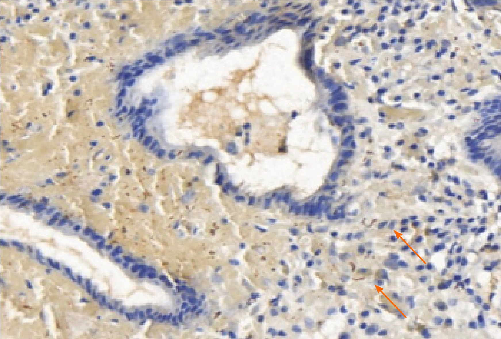
Figure 8 Numerous spirochetes were observed by immunohistochemical staining using a monoclonal antibody against Treponemapallidum (orange arrows, original magnification 400 ×).
- Citation: Lan YM, Yang SW, Dai MG, Ye B, He FY. Gastric syphilis mimicking gastric cancer: A case report. World J Clin Cases 2021; 9(26): 7798-7804
- URL: https://www.wjgnet.com/2307-8960/full/v9/i26/7798.htm
- DOI: https://dx.doi.org/10.12998/wjcc.v9.i26.7798


