Copyright
©The Author(s) 2021.
World J Clin Cases. Sep 16, 2021; 9(26): 7614-7619
Published online Sep 16, 2021. doi: 10.12998/wjcc.v9.i26.7614
Published online Sep 16, 2021. doi: 10.12998/wjcc.v9.i26.7614
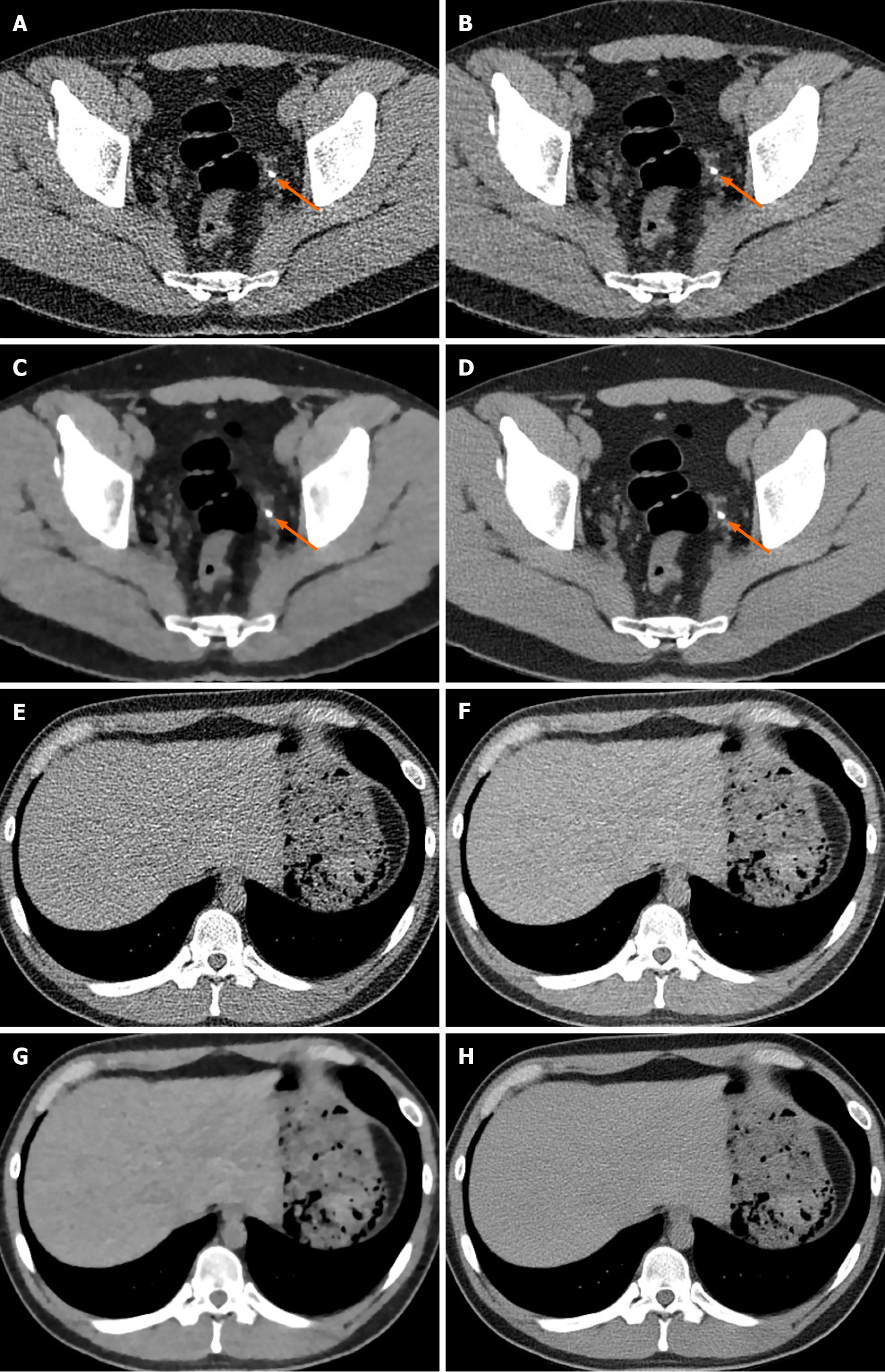
Figure 1 Representative low-dose computed tomography images at 100 kV and 30 mAs (dose length product, approximately 93.
4 mGy· cm; effective radiation dose, approximately 1.401 mSv) using four image reconstruction techniques: Filtered back projection; iDose4, hybrid iterative reconstruction; iterative model reconstruction, fully iterative reconstruction; ClariCT, deep learning based image reconstruction. The arrows point to a left distal ureter stone of 5-mm diameter. The noise in the images produced by FBP (A, E), iDose4 (B, F), IMR (C, G) and ClariCT (D, H) is decreased and the image quality, improved. An unfamiliar plastic-looking noise texture is observed in the high-level iterative reconstruction (C, G). On the other hand, deep learning-based image reconstruction (D, H) sharpened structural margins and produced favorable images.
- Citation: Park SB. Advances in deep learning for computed tomography denoising. World J Clin Cases 2021; 9(26): 7614-7619
- URL: https://www.wjgnet.com/2307-8960/full/v9/i26/7614.htm
- DOI: https://dx.doi.org/10.12998/wjcc.v9.i26.7614


