Copyright
©The Author(s) 2021.
World J Clin Cases. Sep 6, 2021; 9(25): 7551-7557
Published online Sep 6, 2021. doi: 10.12998/wjcc.v9.i25.7551
Published online Sep 6, 2021. doi: 10.12998/wjcc.v9.i25.7551
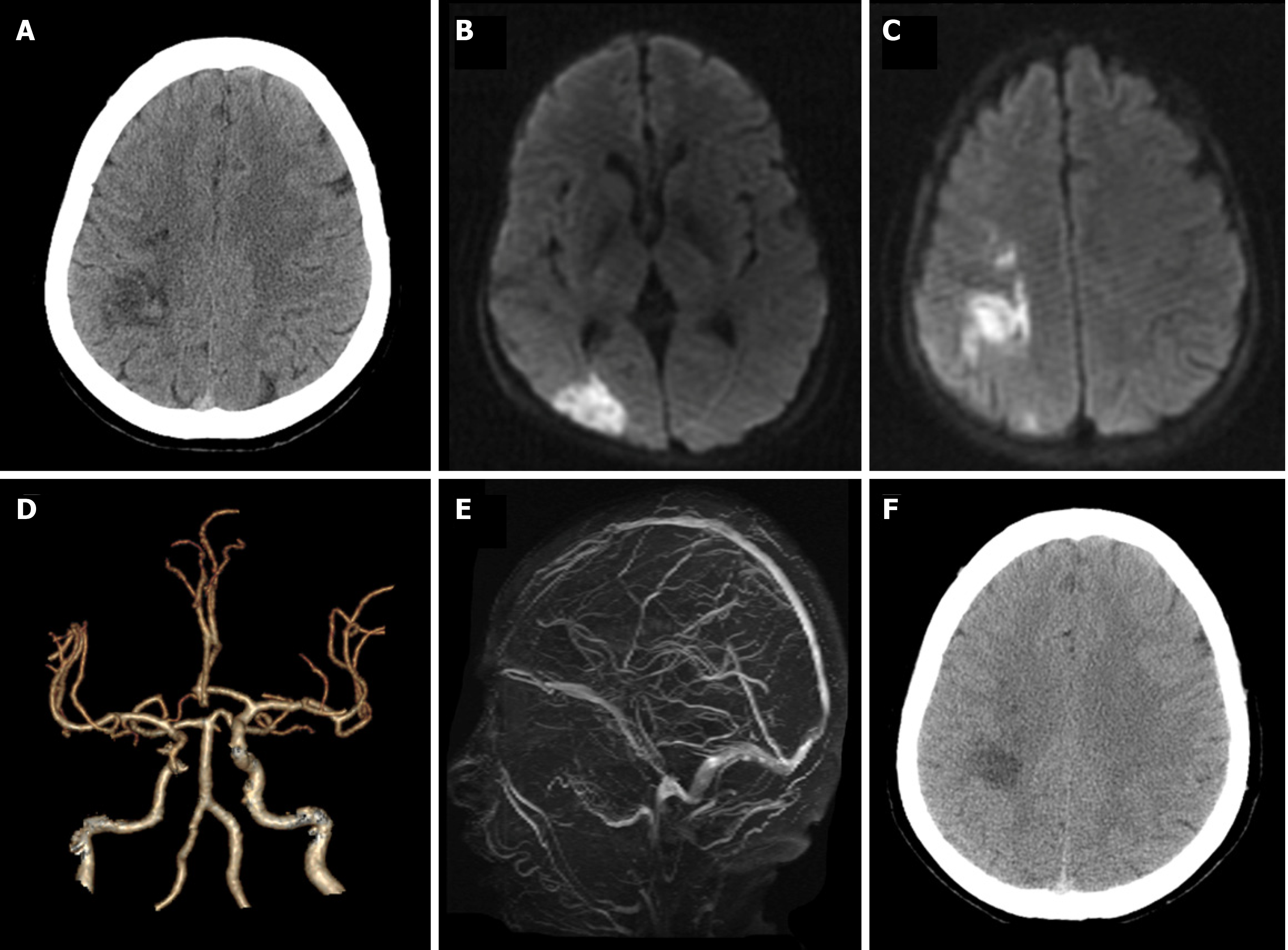
Figure 1 A 57-year-old woman with hemorrhagic infarction due to polycythemia vera.
A: Computed tomography (CT) showed patchy high-density changes in right parietal low-density lesions; B and C: Diffusion-weighted imaging showed acute ischemia in the right parietal and occipital lobes; D: CT-angiography showed mild atherosclerosis; E: Magnetic resonance venogram was unremarkable; F: CT re-examination after 1 wk demonstrated the resolution of hemorrhage within the infarction lesion.
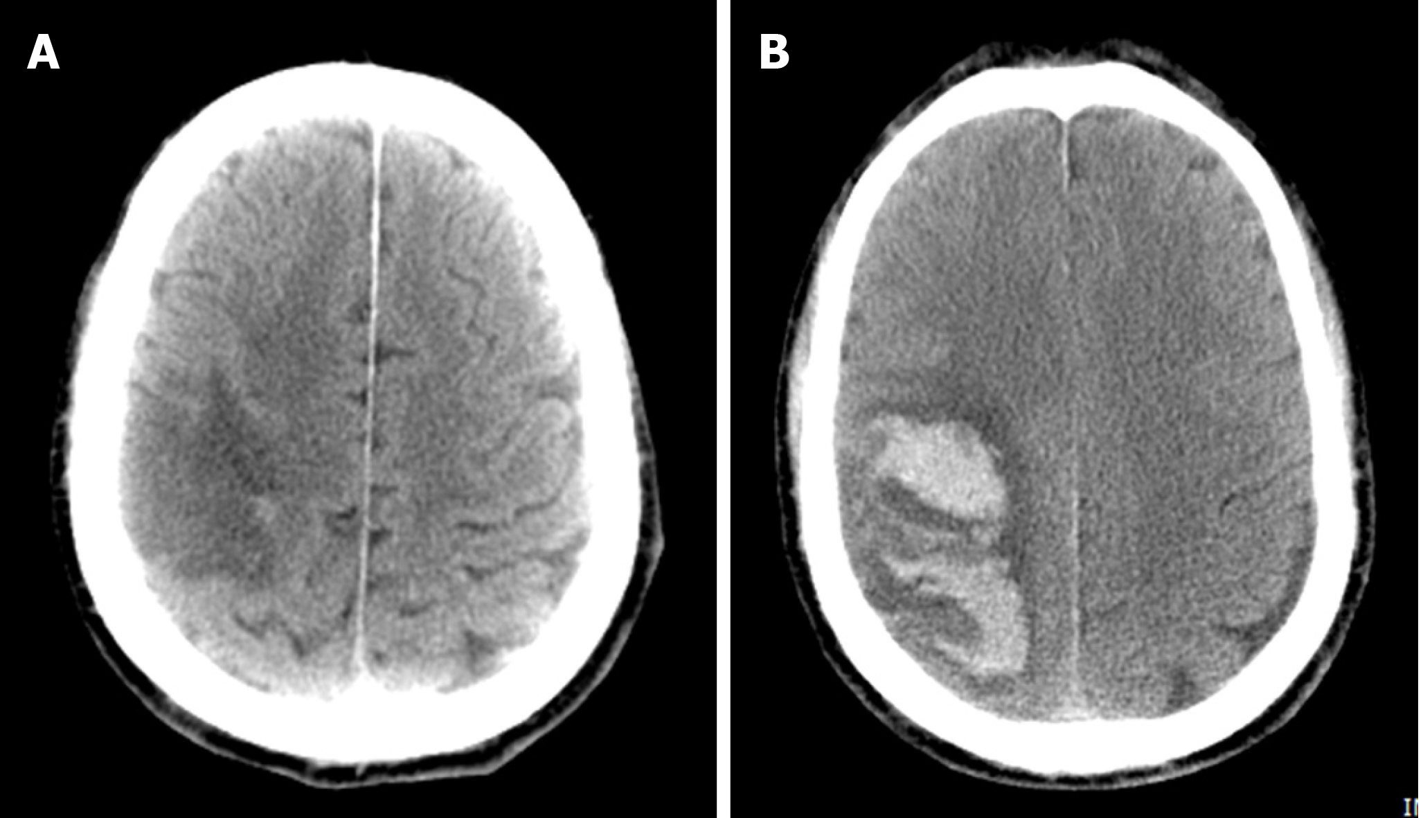
Figure 2 A 68-year-old man with parenchymal hematomas after acute ischemic stroke caused by polycythemia vera.
A: Computed tomography (CT) revealed right parietal and temporal lobe infarction; B: CT re-examination after 3 d revealed a hematoma within the infarct lesion of the right cerebral hemisphere.
- Citation: Cao YY, Cao J, Bi ZJ, Xu SB, Liu CC. Hemorrhagic transformation after acute ischemic stroke caused by polycythemia vera: Report of two case. World J Clin Cases 2021; 9(25): 7551-7557
- URL: https://www.wjgnet.com/2307-8960/full/v9/i25/7551.htm
- DOI: https://dx.doi.org/10.12998/wjcc.v9.i25.7551


