Copyright
©The Author(s) 2021.
World J Clin Cases. Sep 6, 2021; 9(25): 7484-7489
Published online Sep 6, 2021. doi: 10.12998/wjcc.v9.i25.7484
Published online Sep 6, 2021. doi: 10.12998/wjcc.v9.i25.7484
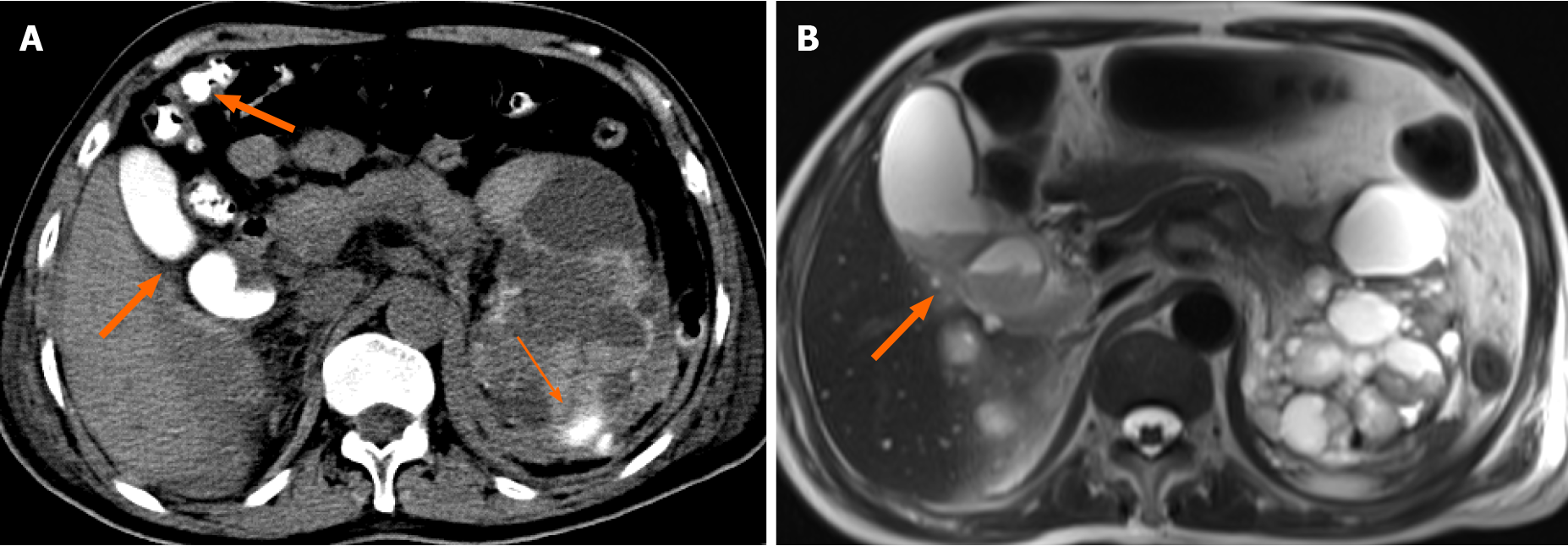
Figure 1 Abdominal computed tomography and magnetic resonance imaging of the patient after renal artery embolization.
A: High density in the gallbladder and colon was vicarious excretion of contrast medium (CM) (thick arrow), while in the left kidney CM had spilled out of the renal artery (thin arrow); B: T2-weighted image shows that the low signal in the gallbladder was sludge (thick arrow).
- Citation: Han ZH, He ZM, Chen WH, Wang CY, Wang Q. Octreotide-induced acute life-threatening gallstones after vicarious contrast medium excretion: A case report. World J Clin Cases 2021; 9(25): 7484-7489
- URL: https://www.wjgnet.com/2307-8960/full/v9/i25/7484.htm
- DOI: https://dx.doi.org/10.12998/wjcc.v9.i25.7484


