Copyright
©The Author(s) 2021.
World J Clin Cases. Sep 6, 2021; 9(25): 7297-7305
Published online Sep 6, 2021. doi: 10.12998/wjcc.v9.i25.7297
Published online Sep 6, 2021. doi: 10.12998/wjcc.v9.i25.7297
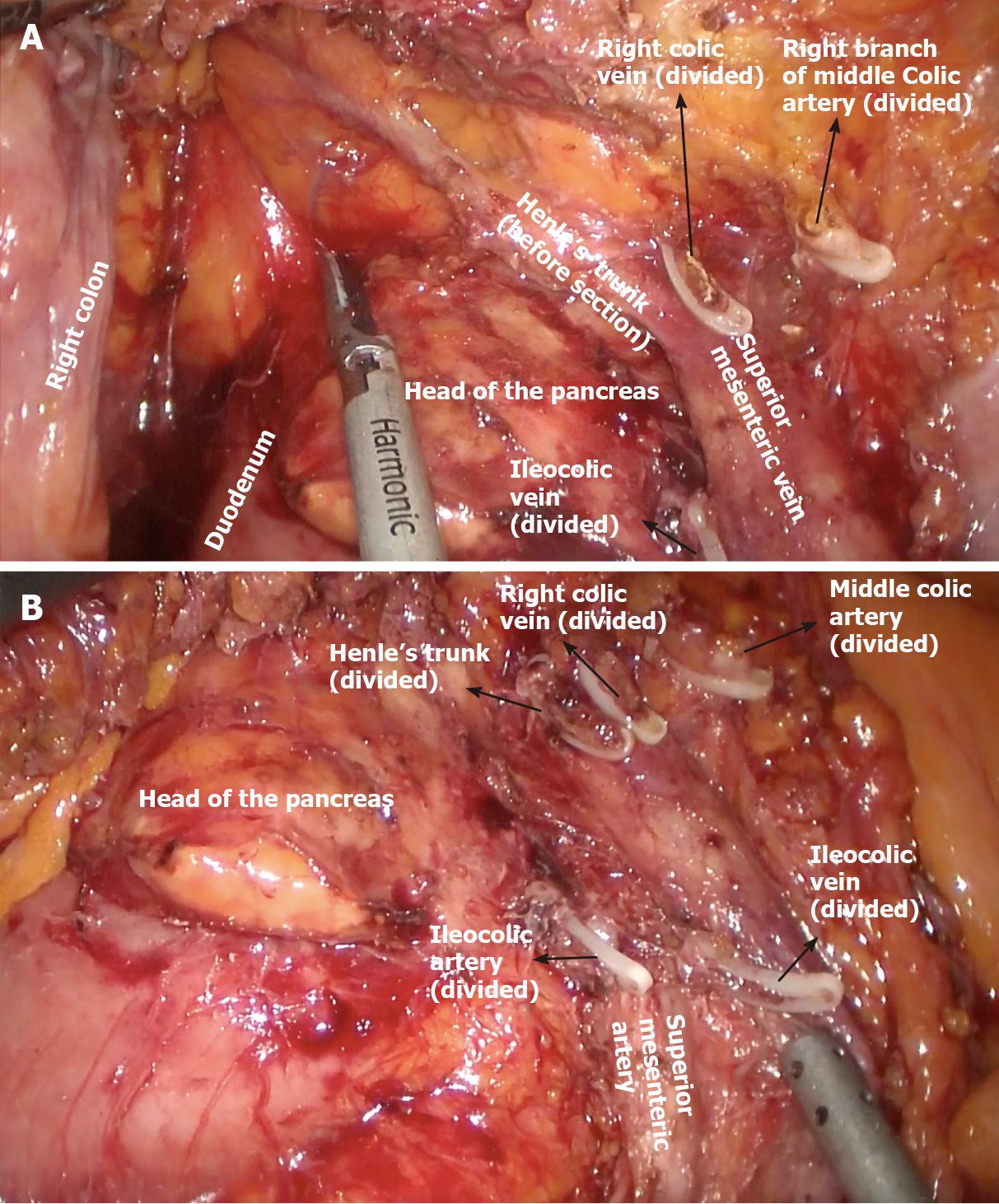
Figure 1 Complete mesocolic excision during right colectomy.
A: Lymphoadipose tissue covering the head of the pancreas after sectioning of the superior right colic vein (SRCV) at its confluence in the gastrocolic trunk of Henle (before dissection), and right branch of the middle colic artery; B: Lympho
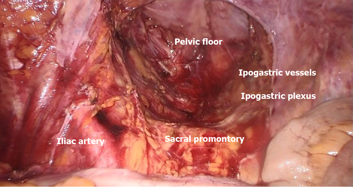
Figure 2
Surgical field after total mesorectal excision dissection in low rectal cancer (Courtesy of Professor Giuseppe Sica, Tor Vergata University of Rome).
- Citation: Franceschilli M, Di Carlo S, Vinci D, Sensi B, Siragusa L, Bellato V, Caronna R, Rossi P, Cavallaro G, Guida A, Sibio S. Complete mesocolic excision and central vascular ligation in colorectal cancer in the era of minimally invasive surgery. World J Clin Cases 2021; 9(25): 7297-7305
- URL: https://www.wjgnet.com/2307-8960/full/v9/i25/7297.htm
- DOI: https://dx.doi.org/10.12998/wjcc.v9.i25.7297


