Copyright
©The Author(s) 2021.
World J Clin Cases. Aug 26, 2021; 9(24): 7205-7211
Published online Aug 26, 2021. doi: 10.12998/wjcc.v9.i24.7205
Published online Aug 26, 2021. doi: 10.12998/wjcc.v9.i24.7205
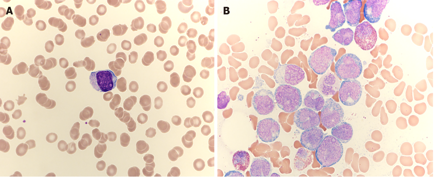
Figure 1 Blood smear and bone marrow aspiration results.
A: The blood smear shows 44% blast cells (× 1000); B: The bone marrow aspiration showed 77.3% myoblasts (× 1000).
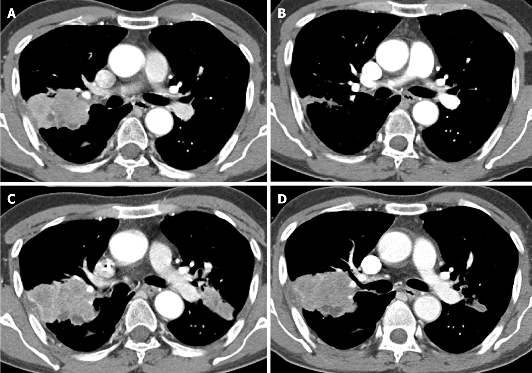
Figure 2 Contrast-enhanced chest computed tomography findings.
A: Before erlotinib treatment, a 59 mm × 42 mm heterogenous enhancing lobulated mass in the right upper lobe (RUL) is closely abutting the adjacent costal pleura with focal thickening and retraction; B: During erlotinib treatment, the mass in the RUL is markedly improved; C: Four months after discontinuing the erlotinib due to leukemia induction chemotherapy, the mass in the RUL is enlarged, and a spiculated mass is visible in the left upper lobe (LUL) (24 mm × 17 mm); D: Two months after pemetrexed/cisplatin treatment, no remarkable changes in the RUL and LUL masses are noted.
- Citation: Koo SM, Kim KU, Kim YK, Uh ST. Therapy-related myeloid leukemia during erlotinib treatment in a non-small cell lung cancer patient: A case report. World J Clin Cases 2021; 9(24): 7205-7211
- URL: https://www.wjgnet.com/2307-8960/full/v9/i24/7205.htm
- DOI: https://dx.doi.org/10.12998/wjcc.v9.i24.7205


