Copyright
©The Author(s) 2021.
World J Clin Cases. Aug 26, 2021; 9(24): 7181-7188
Published online Aug 26, 2021. doi: 10.12998/wjcc.v9.i24.7181
Published online Aug 26, 2021. doi: 10.12998/wjcc.v9.i24.7181
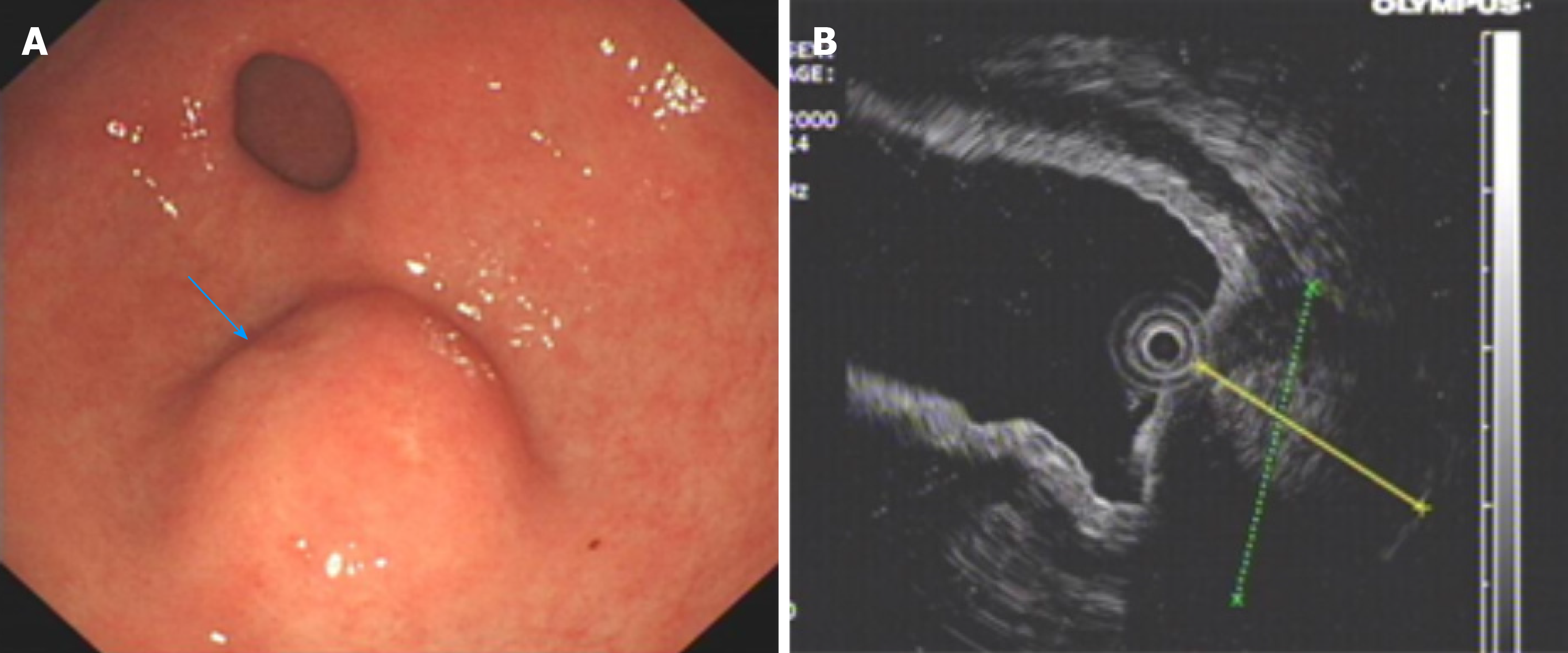
Figure 1 Preoperative endoscopy and endoscopic ultrasonography.
A: Endoscopy of the upper digestive tract revealed a submucosal tumor of great curvature in the gastric antrum; B: Endoscopic ultrasonography identified a 2.4 cm × 1.8 cm lump located in the gastric antrum originating from the muscularis propria with both intraluminal and extracavity growth.
Figure 2 Computed tomography plain scan of gastrointestinal stromal tumors and gastric glomus tumor.
A: Gastric glomus tumor presented as submucosal masses with clear boundaries and uniform density (orange arrow); B: Plain computed tomography scan showed that the gastrointestinal stromal tumors were nodular (blue arrow).
Figure 3 The arterial period of gastrointestinal stromal tumors and gastric glomus tumor.
A: Enhanced computed tomography showed that the arterial phase of gastrointestinal stromal tumors presented mild heterogeneous enhancement (blue arrow); B: The arterial phase of gastric glomus tumor presented significant enhancement (orange arrow).
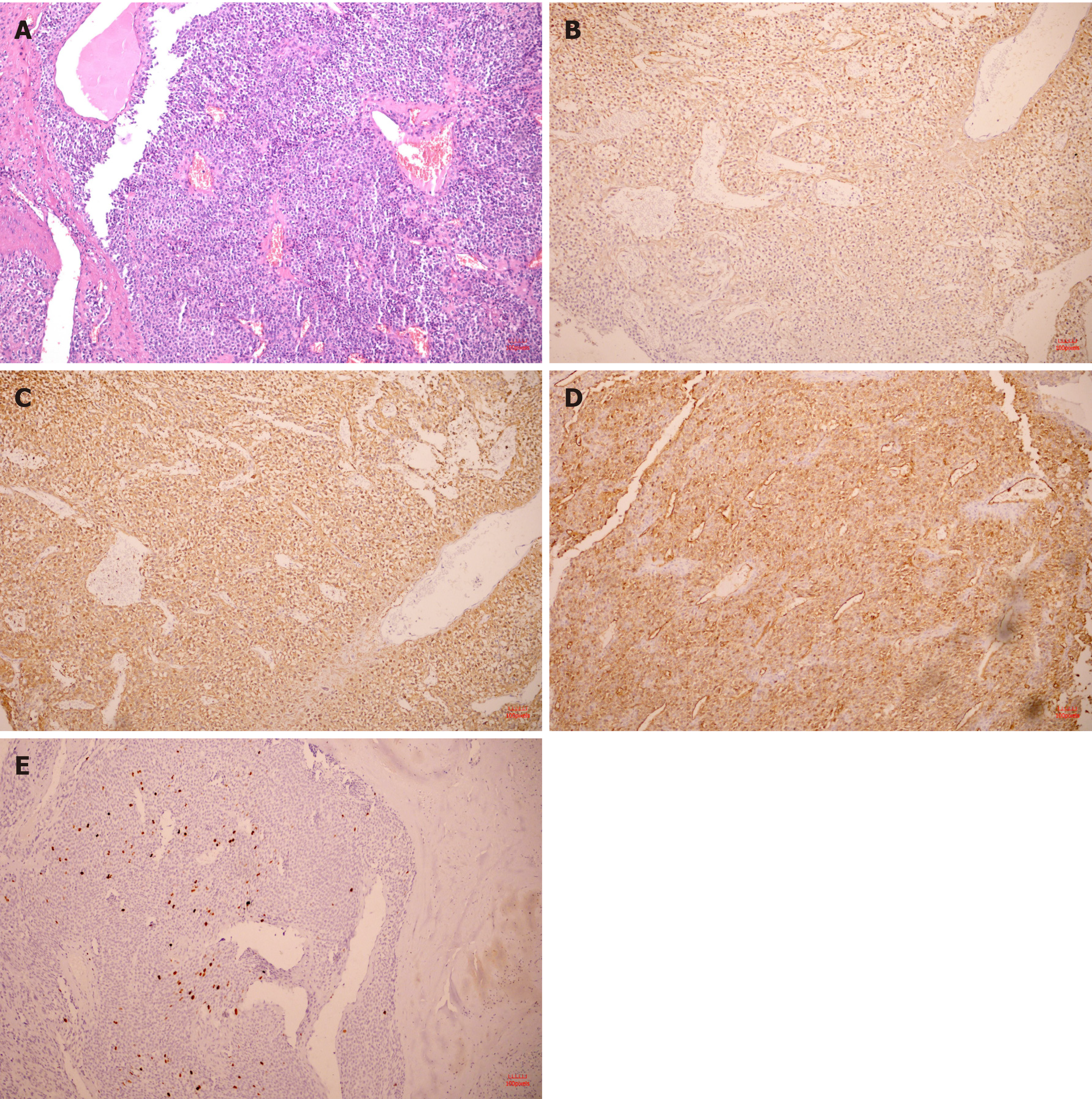
Figure 4 Hematoxylin and eosin pathological sections and immunohistochemistry after operation.
A: The tumor was composed of aggregated round and fusiform cells surrounded by capillaries (10 ×); B: Immunohistochemistry showed spinal muscular atrophy (+, 10 ×); C: H-caldesmon (+, 10 ×); D: cluster of differentiation 34 (+, 10 ×); E: A Ki-67 positive rate of 2% (10 ×).
Figure 5 Cutting the mucosa to the lamina propria using a dual knife.
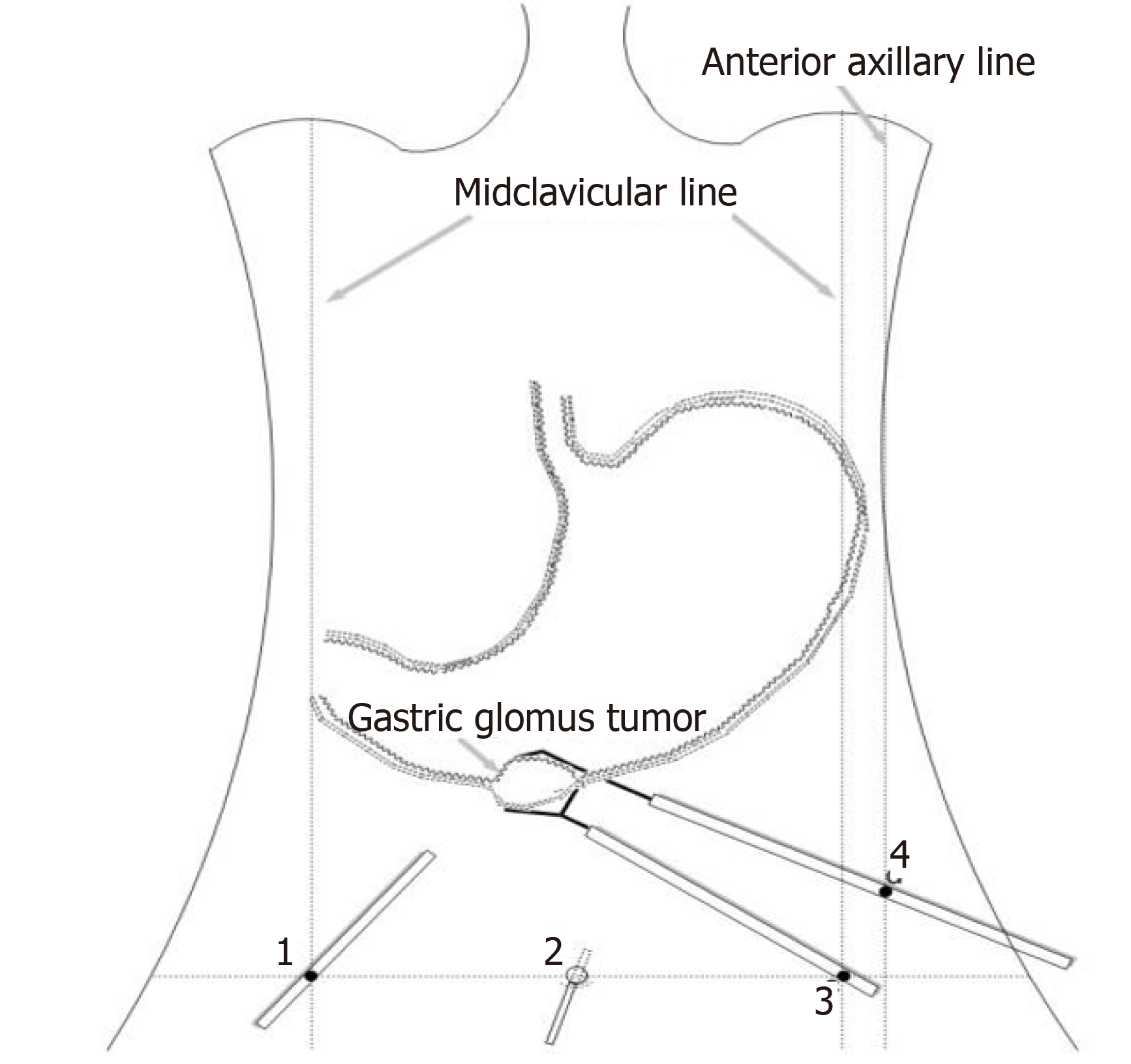
Figure 6 Laparoscopic-assisted resection of the tumor.
Figure 7 The delay period of gastrointestinal stromal tumors and gastric glomus tumor.
Gastric glomus tumor (orange arrow) was more obvious than gastrointestinal stromal tumors (blue arrow) during the delay period. A: Gastrointestinal stromal tumors; B: Gastric glomus tumor.
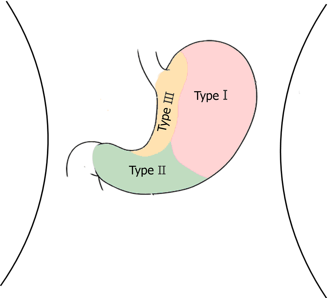
Figure 8 Tumor location and corresponding traditional surgical technique.
Type I: Laparoscopic partial gastrectomy; Type II: Laparoscopic distal gastrectomy; Type III: Laparoscopic transgastric resection.
- Citation: Wang WH, Shen TT, Gao ZX, Zhang X, Zhai ZH, Li YL. Combined laparoscopic-endoscopic approach for gastric glomus tumor: A case report . World J Clin Cases 2021; 9(24): 7181-7188
- URL: https://www.wjgnet.com/2307-8960/full/v9/i24/7181.htm
- DOI: https://dx.doi.org/10.12998/wjcc.v9.i24.7181


