Copyright
©The Author(s) 2021.
World J Clin Cases. Aug 26, 2021; 9(24): 7085-7091
Published online Aug 26, 2021. doi: 10.12998/wjcc.v9.i24.7085
Published online Aug 26, 2021. doi: 10.12998/wjcc.v9.i24.7085
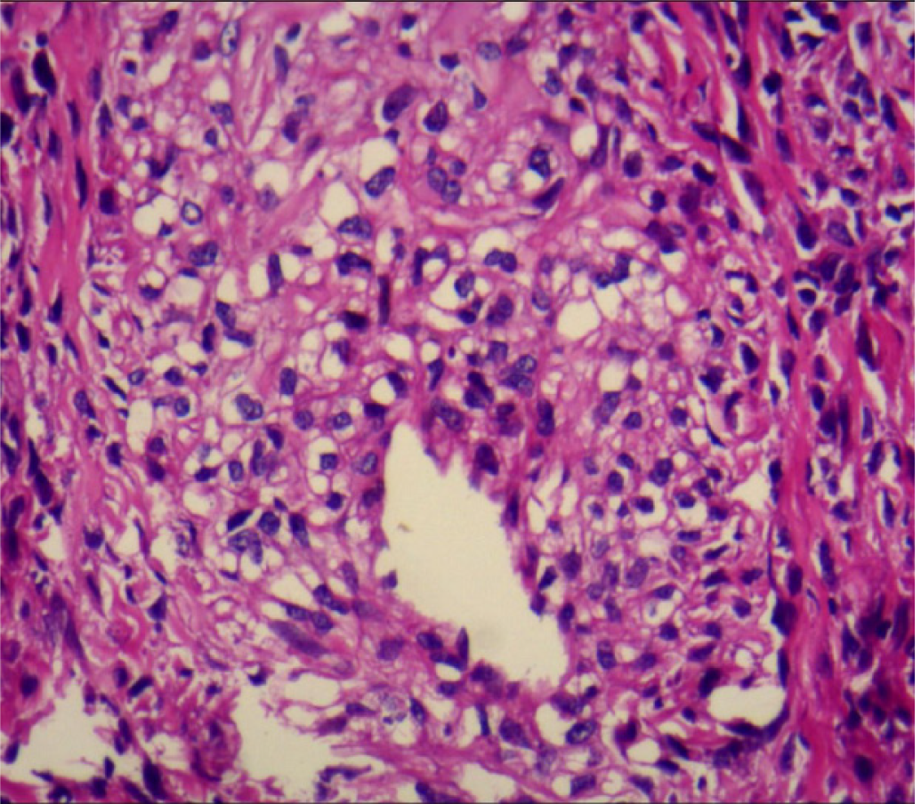
Figure 1 Pathology of the renal angioleiomyolipoma (hematoxylin-eosin staining).
The patient had multiple renal angioleiomyolipomas excised in 2002 and 2008. Magnification: × 800.
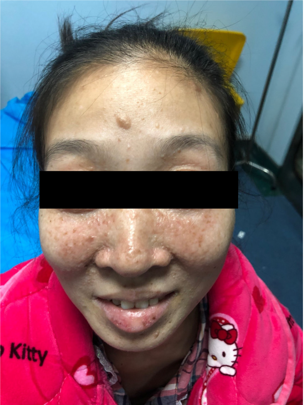
Figure 2 Facial angiofibroma developed after the patient was pregnant in 2013.
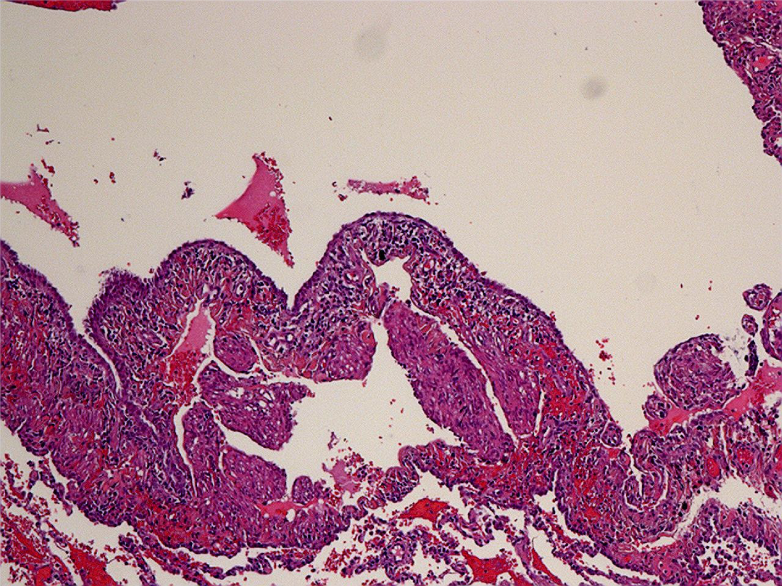
Figure 3 Pathology of lung lymphangioleiomyomatosis (hematoxylin-eosin staining).
Magnification: × 200.
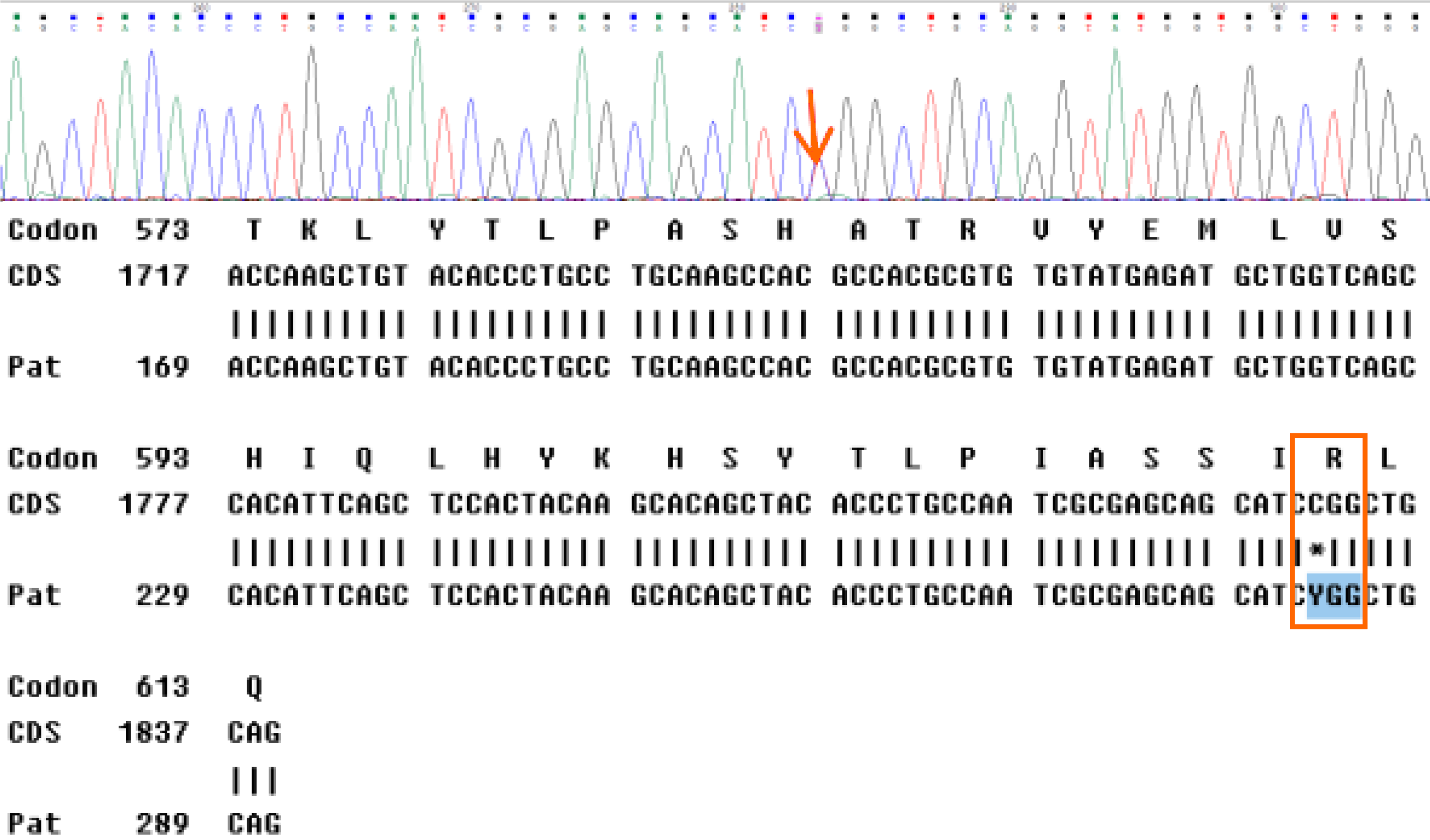
Figure 4 High-throughput sequencing of peripheral blood DNA.
The heterozygous mutation c.1831C>T (p.Arg611Trp) in the TSC2 gene was confirmed.
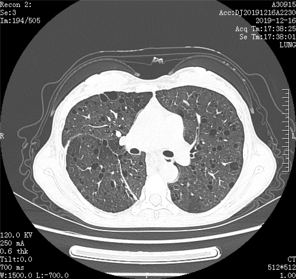
Figure 5 High-resolution chest computed tomography.
Computed tomography revealed interstitial changes, multiple lytic and lucent lesions of varying sizes, bilateral pulmonary nodules, and multiple fat density areas in the inferior mediastinum.
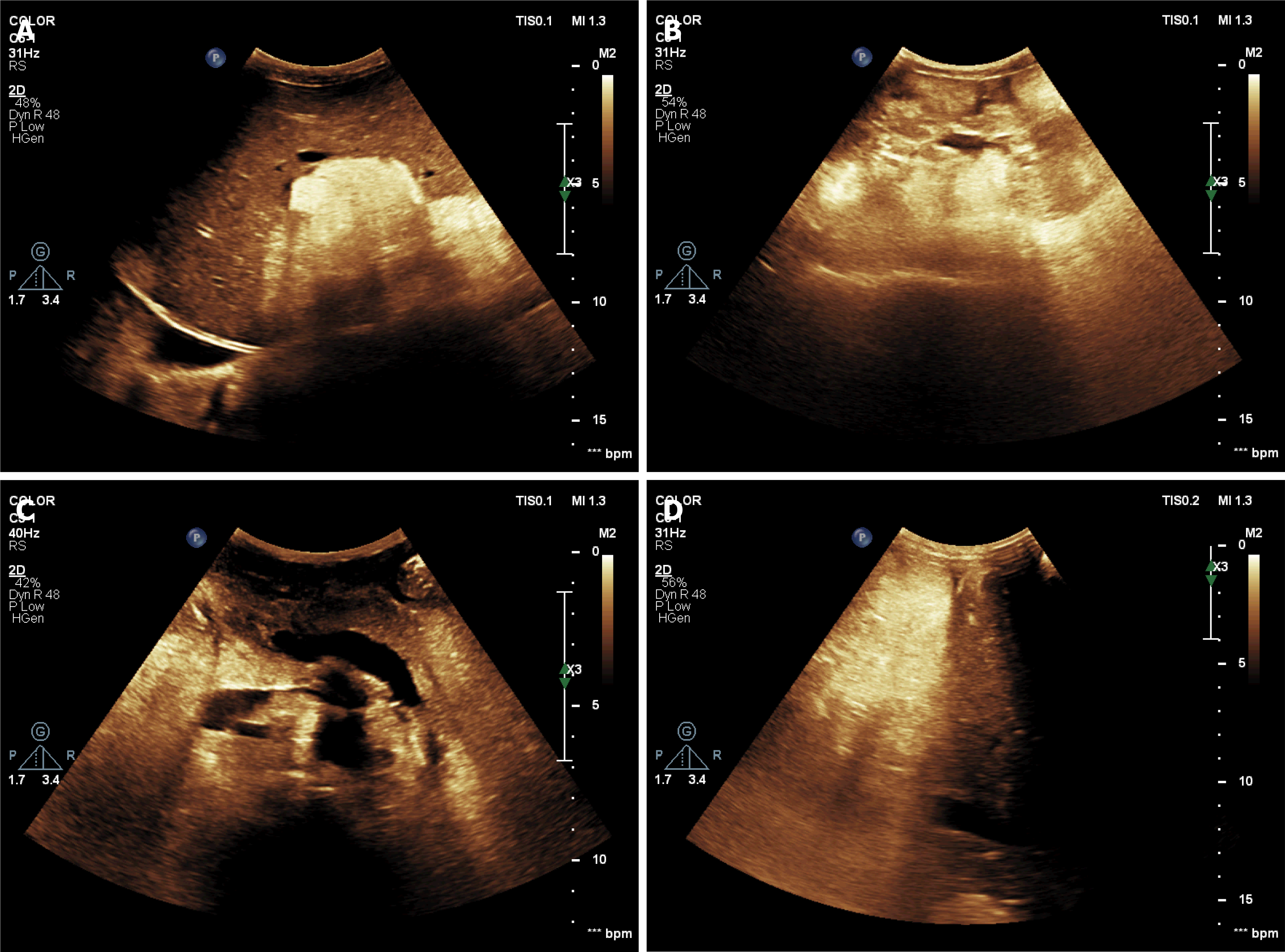
Figure 6 Ultrasonography revealed multiple high echogenic masses.
A: Liver; B: Right kidney; C: Retroperitoneum; D: Inferior mediastinum.
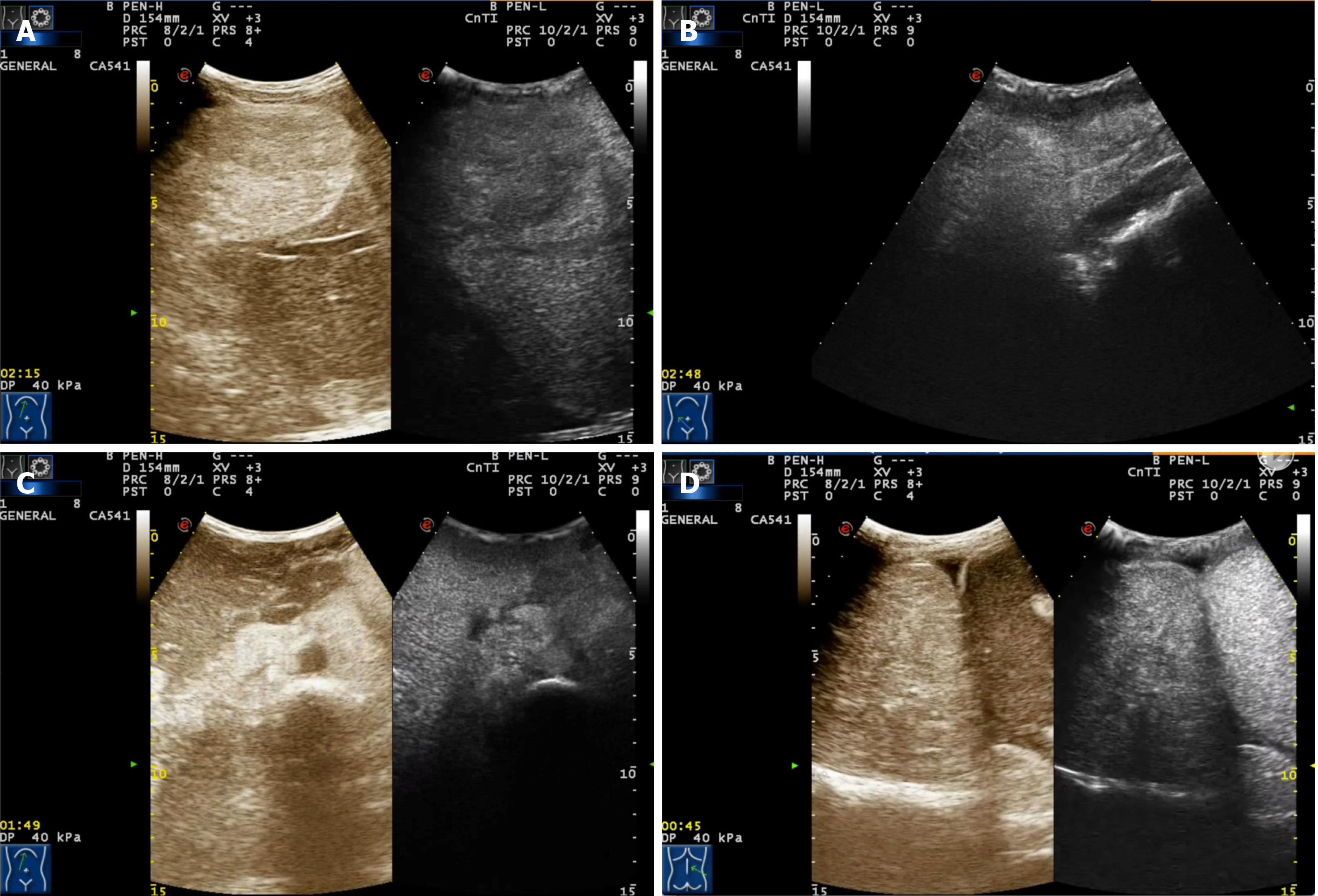
Figure 7 Contrast-enhanced ultrasonography revealed high enhancement of the masses.
A: Liver; B: Right kidney; C: Retroperitoneum; D: Inferior mediastinum.
- Citation: Chen HB, Xu XH, Yu CG, Wan MT, Feng CL, Zhao ZY, Mei DE, Chen JL. Tuberous sclerosis complex-lymphangioleiomyomatosis involving several visceral organs: A case report. World J Clin Cases 2021; 9(24): 7085-7091
- URL: https://www.wjgnet.com/2307-8960/full/v9/i24/7085.htm
- DOI: https://dx.doi.org/10.12998/wjcc.v9.i24.7085


