Copyright
©The Author(s) 2021.
World J Clin Cases. Aug 16, 2021; 9(23): 6956-6963
Published online Aug 16, 2021. doi: 10.12998/wjcc.v9.i23.6956
Published online Aug 16, 2021. doi: 10.12998/wjcc.v9.i23.6956
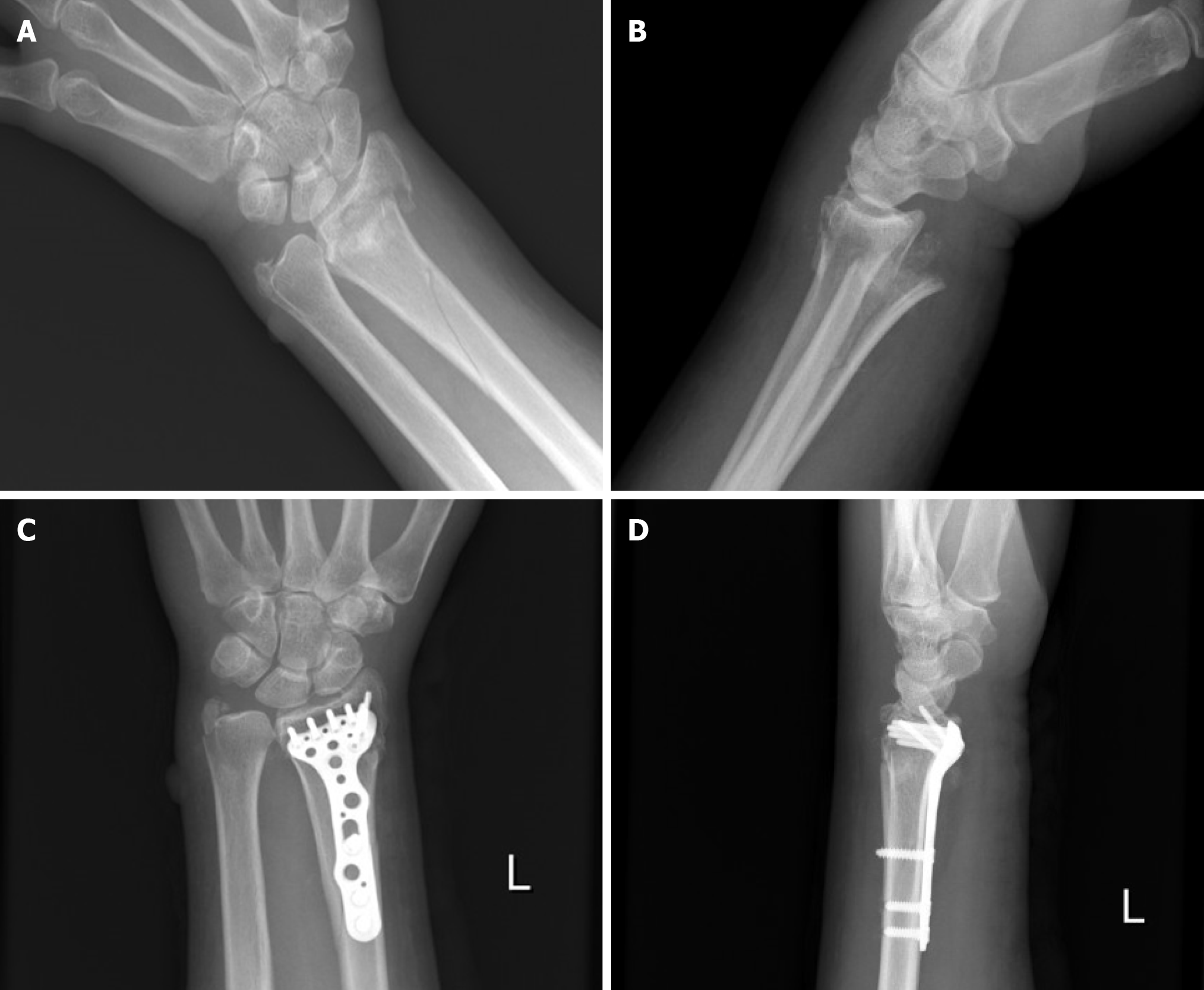
Figure 1 Anterior-posterior and lateral wrist radiography of case 1.
A and B: Anterior-posterior and lateral wrist radiographs revealed a left distal radial fracture involving the radial trunk, accompanied by dorsoradial displacement and an ulnar styloid fracture; C and D: Anterior-posterior and lateral wrist radiography after surgical open reduction and fixation with a volar plate.
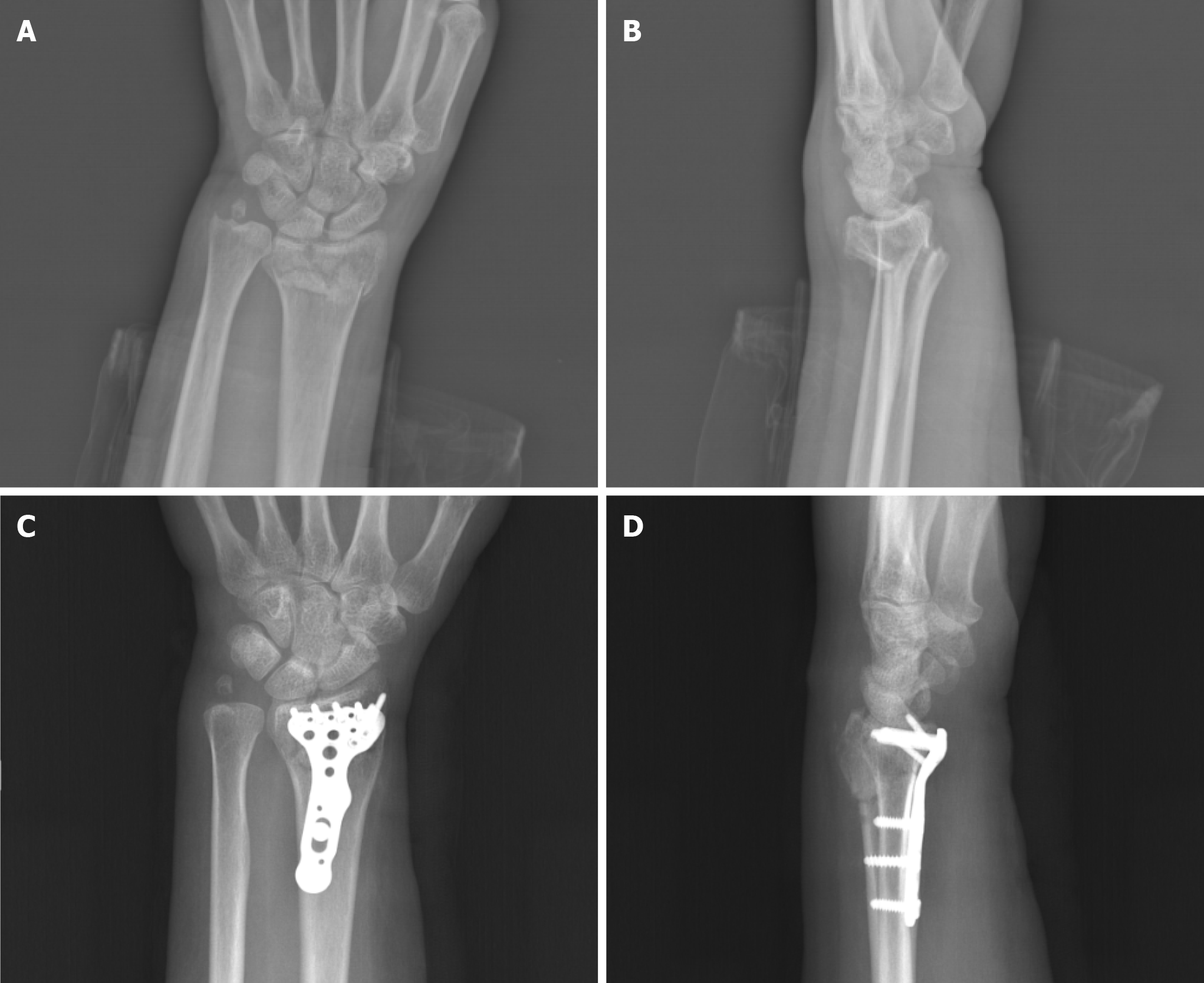
Figure 2 Anterior-posterior and lateral wrist radiography of case 2.
A and B: Anterior-posterior and lateral wrist radiographs revealed a left distal radial intra-articular fracture, accompanied by dorsoradial displacement and an ulnar styloid fracture; C and D: Anterior–posterior and lateral wrist radiograph after surgical open reduction and fixation with a volar plate.
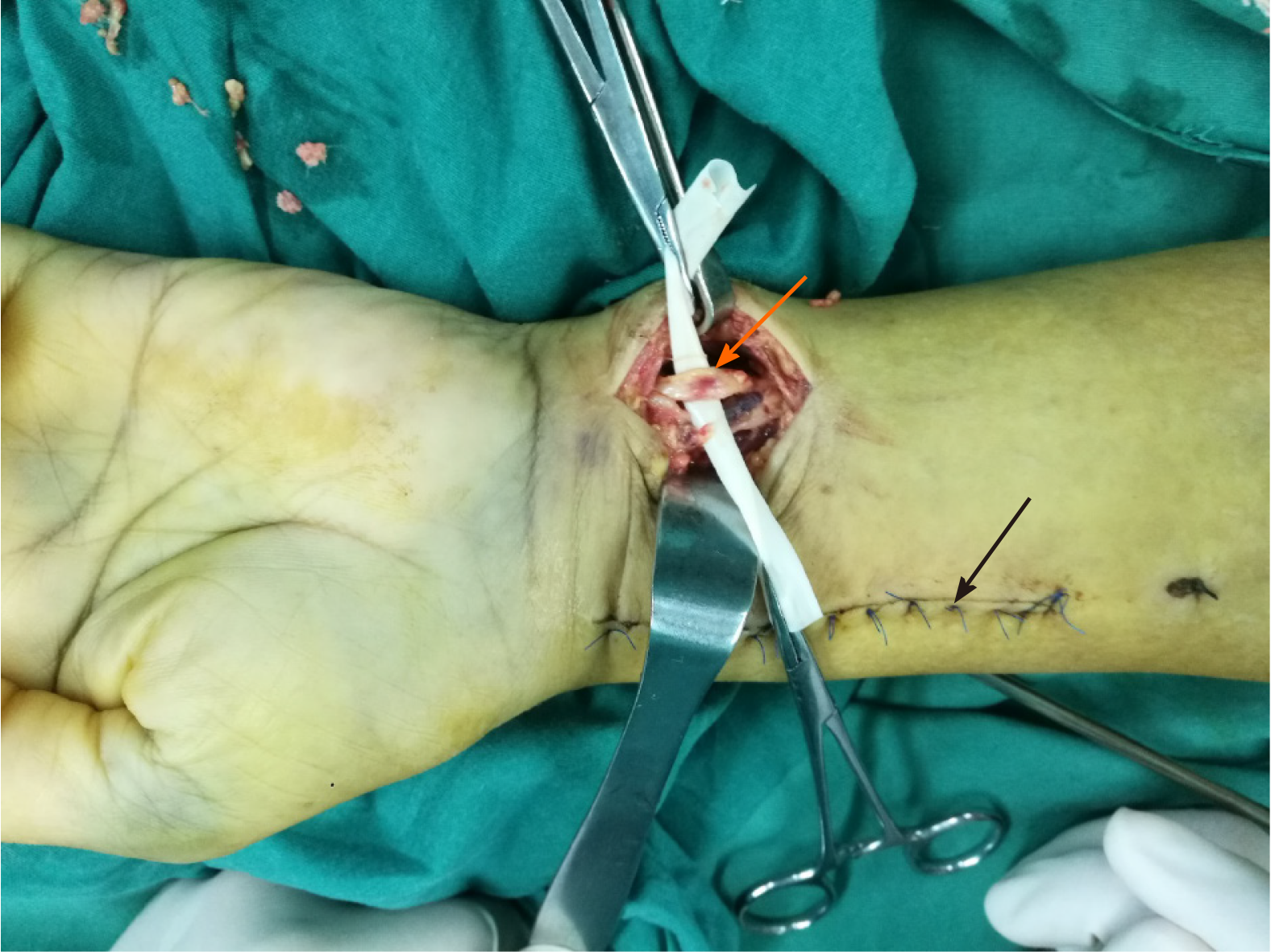
Figure 3 Case 1: Intraoperative photograph showed contusion and swelling of the ulnar nerve (orange arrow) and her first surgical (5 d ago) incision (black arrow).
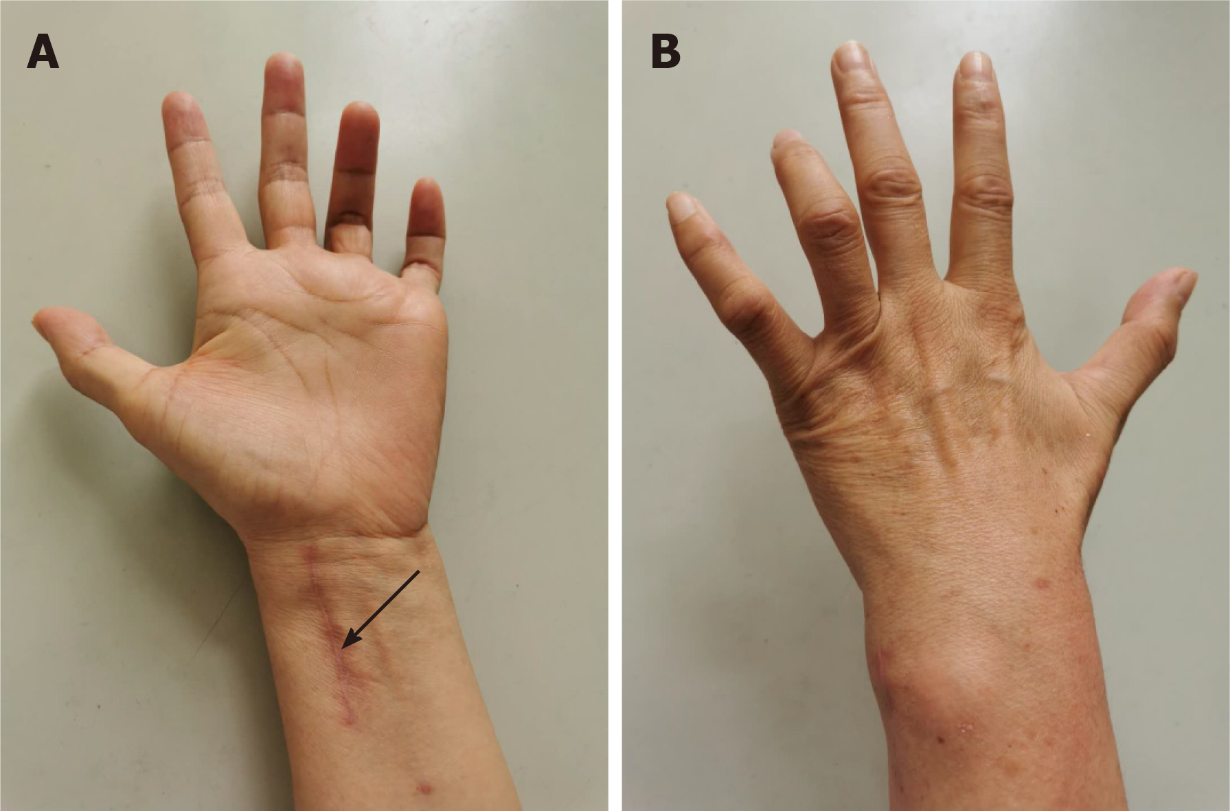
Figure 4 Case 2: At 8 wk after surgery, a typical ulnar claw deformity was noted, and her first surgical incision (10 wk ago) had healed (black arrow).
A: Palmar, B: Dorsal.
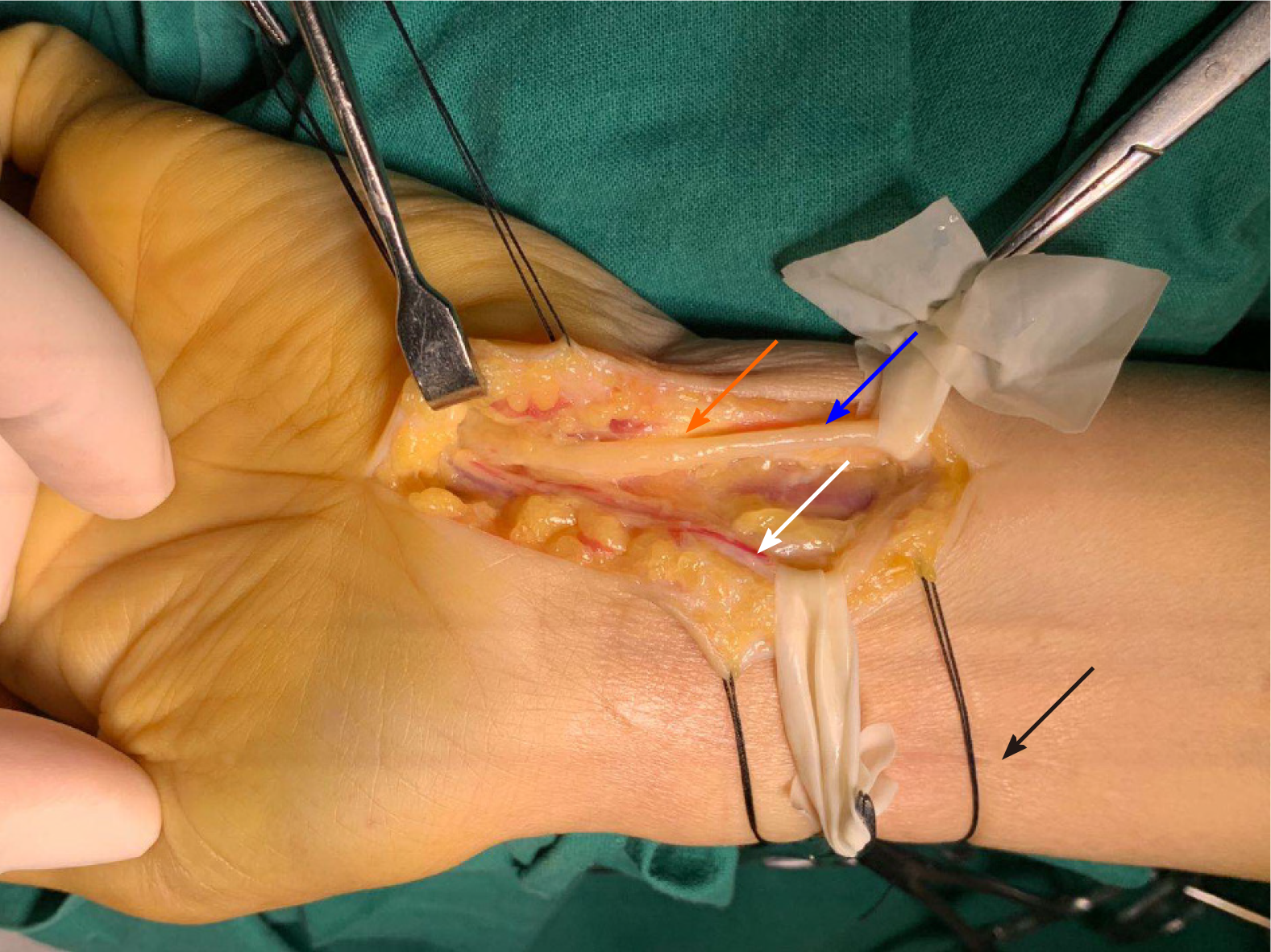
Figure 5 Case 2: Intraoperative photograph shows swelling and compression by the surrounding tissue fibrosis of the ulnar nerve at the level of Guyon’s canal (zone 1).
Swelling ulnar nerve (orange arrow), normal ulnar nerve (blue arrow), ulnar artery (white arrow), first surgical incision (10 wk ago) had healed (black arrow).
- Citation: Yang JJ, Qu W, Wu YX, Jiang HJ. Ulnar nerve injury associated with displaced distal radius fracture: Two case reports. World J Clin Cases 2021; 9(23): 6956-6963
- URL: https://www.wjgnet.com/2307-8960/full/v9/i23/6956.htm
- DOI: https://dx.doi.org/10.12998/wjcc.v9.i23.6956


