Copyright
©The Author(s) 2021.
World J Clin Cases. Aug 16, 2021; 9(23): 6935-6942
Published online Aug 16, 2021. doi: 10.12998/wjcc.v9.i23.6935
Published online Aug 16, 2021. doi: 10.12998/wjcc.v9.i23.6935
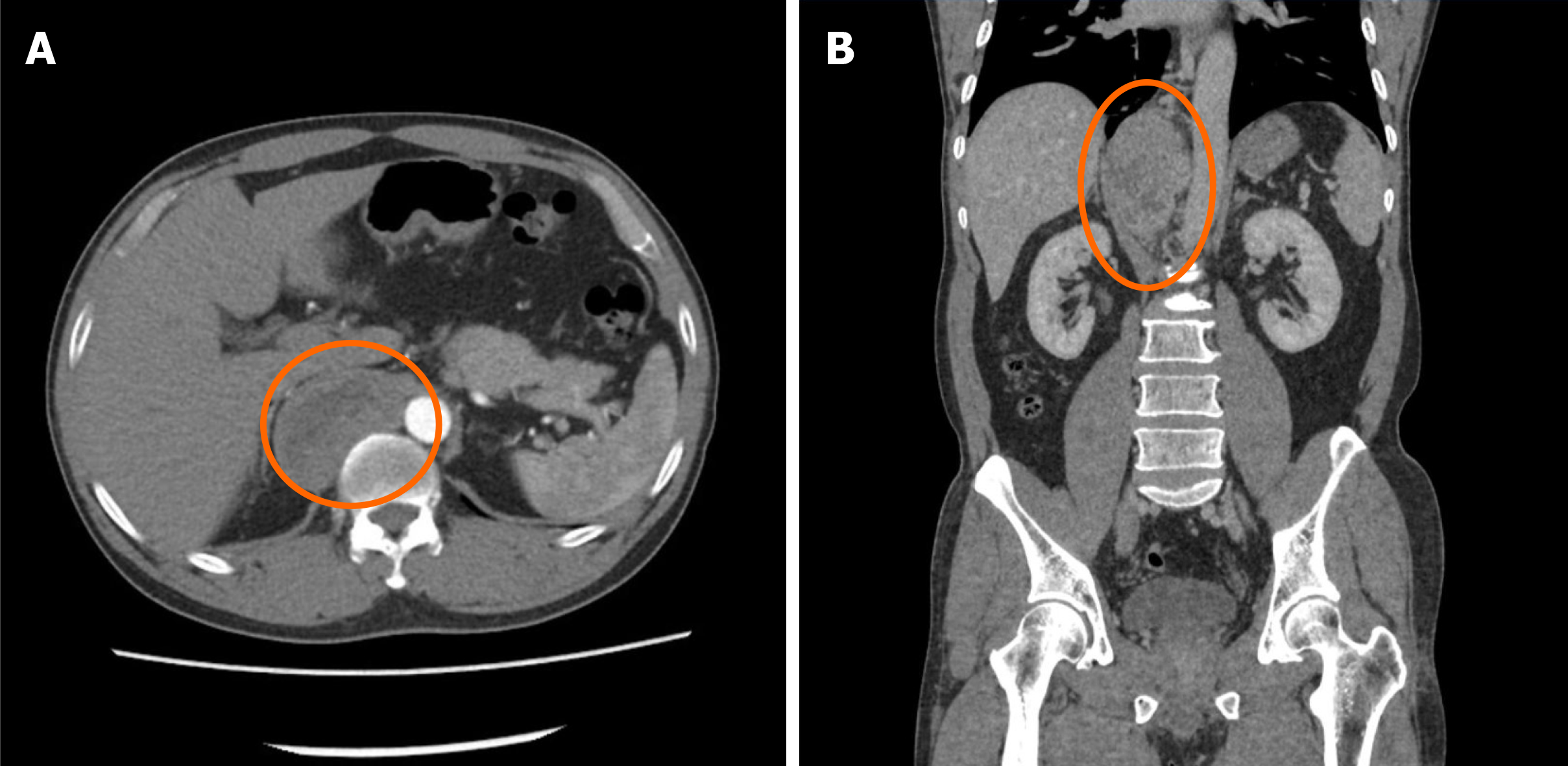
Figure 1 Contrast-enhanced computed tomography + three-dimensional reconstruction of the abdomen and pelvis.
A: Contrast-enhanced computed tomography; B: Three-dimensional reconstruction. At the T10-L1 level of the posterior mediastinal spine on the right anterior side (posterior space of the right phrenic foot), there is an elliptical mixed density shadow, with smooth borders, and the size is about 3.3 cm × 6.9 cm × 8.4 cm.
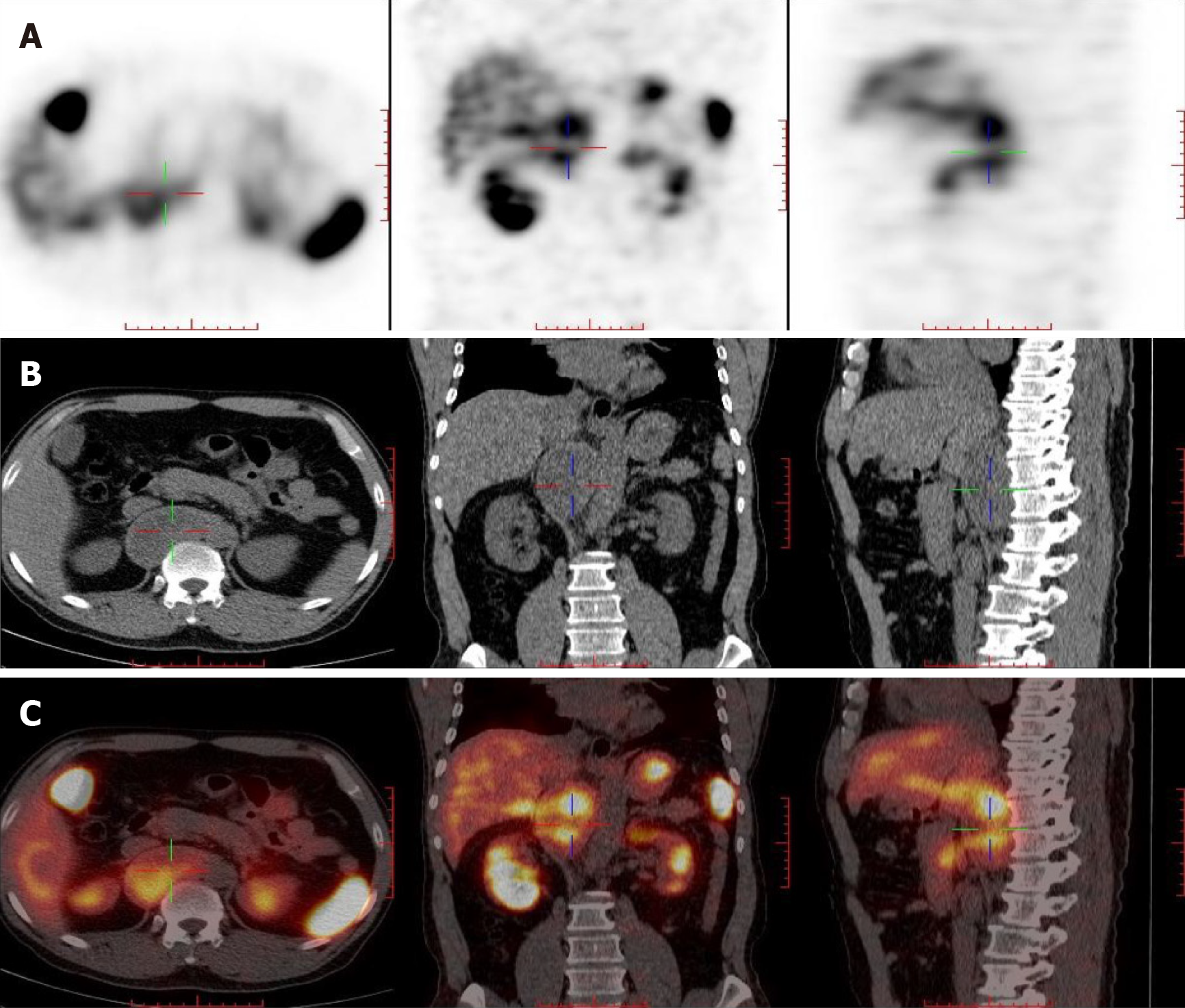
Figure 2 Somatostatin receptor imaging.
A: Contrast agent imaging; B: Computed tomography; C: Fusion imaging of A and B. At the T10-L1 vertebral level on the right side of the abdominal aorta and behind the inferior vena cava, there is a cystic solid space, measuring 7.3 cm × 3.6 cm × 9.4 cm in size, with increased radioactive uptake.
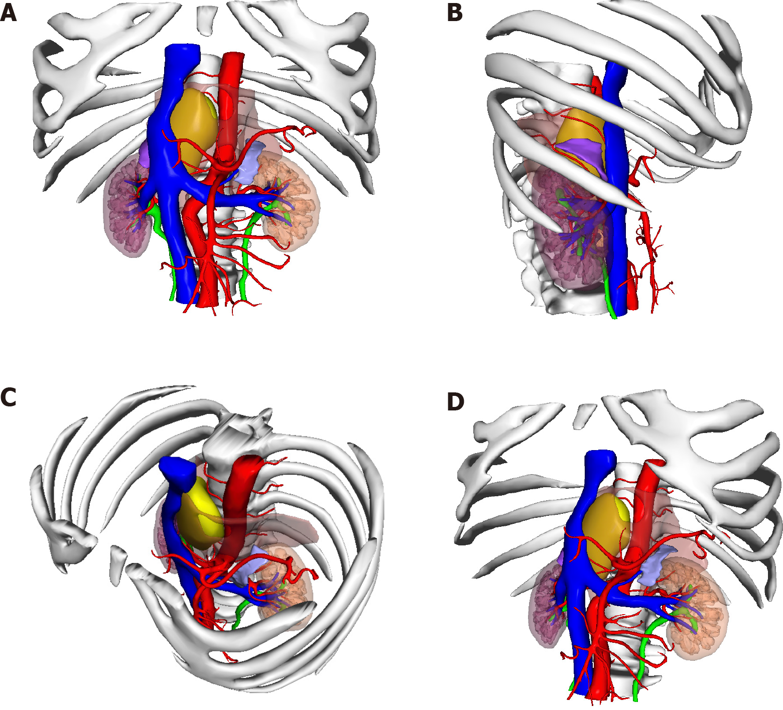
Figure 3 The position of the tumor shown in 3-dimensional printing.
A: Front view; B: Right side view; C: Diagonally above view; D: Diagonally below view. Yellow: Tumor; Orange: Muscle; Blue: Vein; Red: Artery.
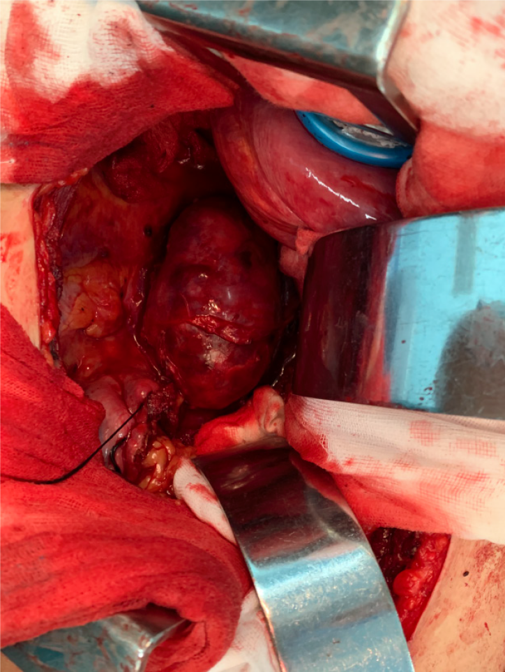
Figure 4 Surgical procedure.
After opening the abdomen, a large retroperitoneal tumor can be seen surrounded by the psoas muscle, with abundant blood supply on the surface.
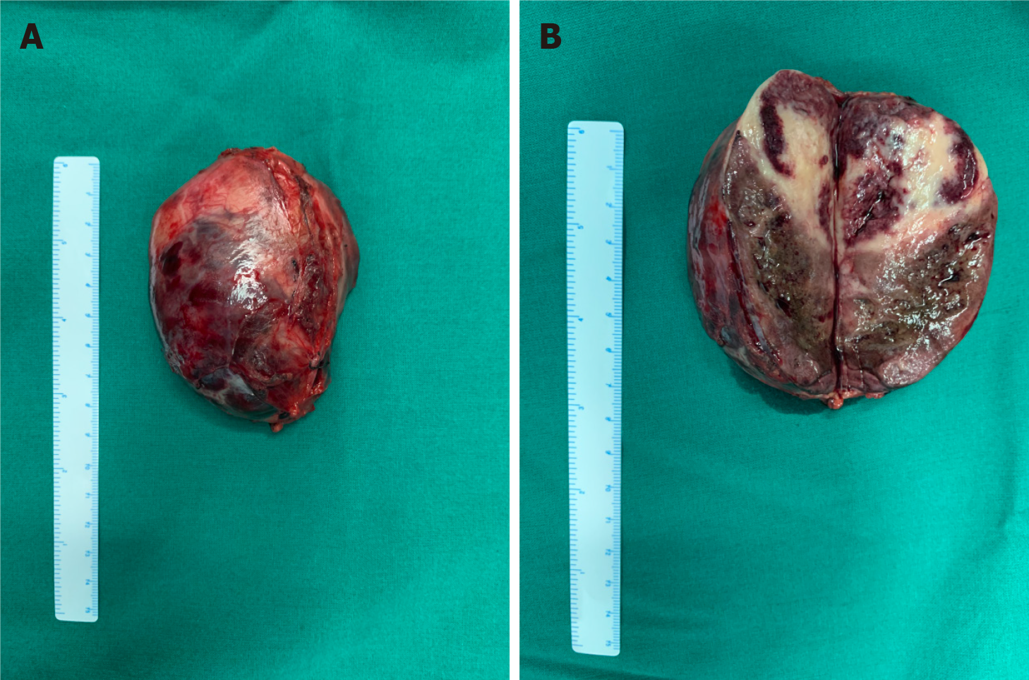
Figure 5 Specimen of a large tumor.
A: Tumor with intact envelope; B: Tumor after incision.
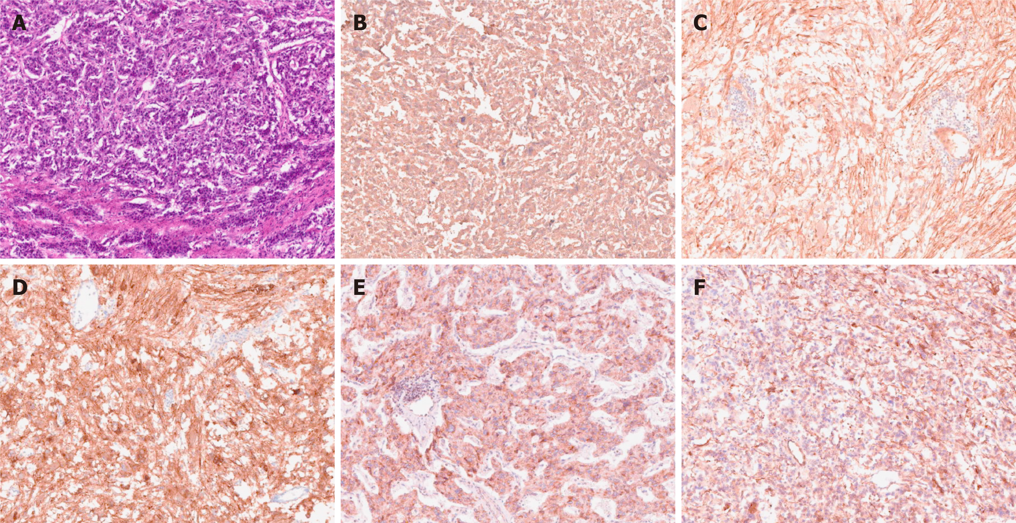
Figure 6 Pathological photos.
Histological examination: A: HE staining; Immunohistochemistry: B: Chromogranin A (+); C: Soluble protein-100 (+); D: Synaptophysin (+); E: Succinate dehydrogenase B (+); F: Neuron specific enolase (+).
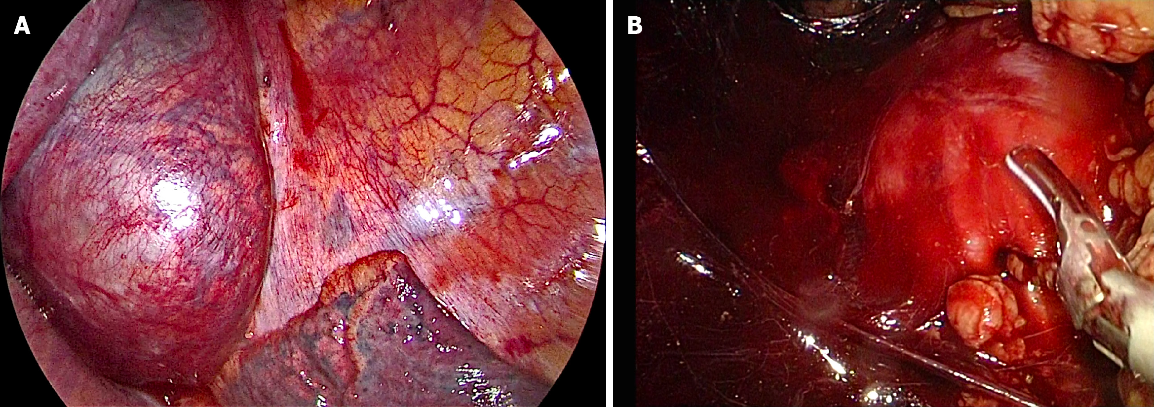
Figure 7 Corresponding images in the process of combined thoracic and laparoscopic detection of the mass.
A: Thoracoscopic image; B: Laparoscopic image.
- Citation: Liu C, Wen J, Li HZ, Ji ZG. Combined thoracoscopic and laparoscopic approach to remove a large retroperitoneal compound paraganglioma: A case report. World J Clin Cases 2021; 9(23): 6935-6942
- URL: https://www.wjgnet.com/2307-8960/full/v9/i23/6935.htm
- DOI: https://dx.doi.org/10.12998/wjcc.v9.i23.6935


