Copyright
©The Author(s) 2021.
World J Clin Cases. Aug 16, 2021; 9(23): 6900-6906
Published online Aug 16, 2021. doi: 10.12998/wjcc.v9.i23.6900
Published online Aug 16, 2021. doi: 10.12998/wjcc.v9.i23.6900
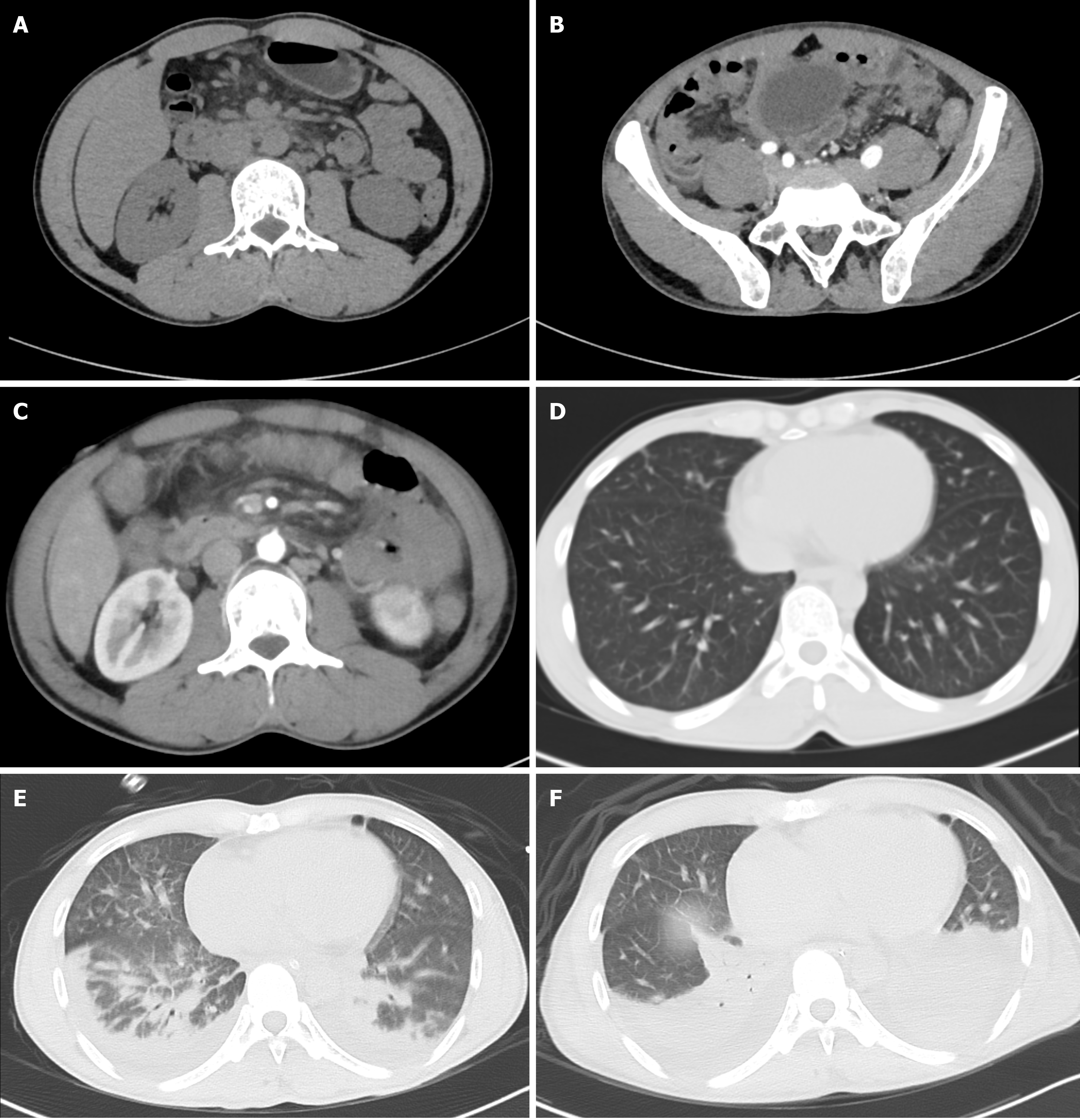
Figure 1 Computed tomography images.
A: Plain computed tomography (CT) scan of the upper abdomen on the first day of hospitalization revealed inflammatory exudation of the peritoneum, with multiple small lymph nodes in the posterior peritoneum; B: Enhanced CT on the second day of hospitalization revealed an enlarged appendix (approximately 9 mm in diameter); C: Enhanced CT on the second day of hospitalization revealed inflammatory exudation of the peritoneum, with multiple small lymph nodes in the posterior peritoneum; D: Plain CT scan on the first day of hospitalization revealed no abnormalities in either lung; E: Enhanced CT on the second day of hospitalization revealed pleural effusion and pulmonary edema in both lungs; F: Plain CT scan on the fourth day of hospitalization revealed expanded lung consolidation range and increased pleural effusion.
Figure 2 Endoscopy on the second day of hospitalization revealed edema of the gastric mucosa but no gastric or duodenal ulcers or perforations.
A: Duodenal bulb; B: Gastric antrum; C: Gastric corpus.
Figure 3 Histopathology of the appendix revealed neutrophil and plasma cell infiltration in the blood vessel wall and around the blood vessel.
A: × 100; B: × 200; C: × 400.
- Citation: Zhou XL, Ye QL, Chen JQ, Li W, Dong HJ. Manifestation of acute peritonitis and pneumonedema in scrub typhus without eschar: A case report. World J Clin Cases 2021; 9(23): 6900-6906
- URL: https://www.wjgnet.com/2307-8960/full/v9/i23/6900.htm
- DOI: https://dx.doi.org/10.12998/wjcc.v9.i23.6900


