Copyright
©The Author(s) 2021.
World J Clin Cases. Aug 16, 2021; 9(23): 6816-6823
Published online Aug 16, 2021. doi: 10.12998/wjcc.v9.i23.6816
Published online Aug 16, 2021. doi: 10.12998/wjcc.v9.i23.6816
Figure 1 Imaging examinations.
A: Dual-source computed tomography scan revealed masses in the right maxillary sinus, nasal cavity and nasopharynx in case 1 (orange arrow); B: Computed tomography scan revealed a mass in the right nasal cavity in case 2 (orange arrow).
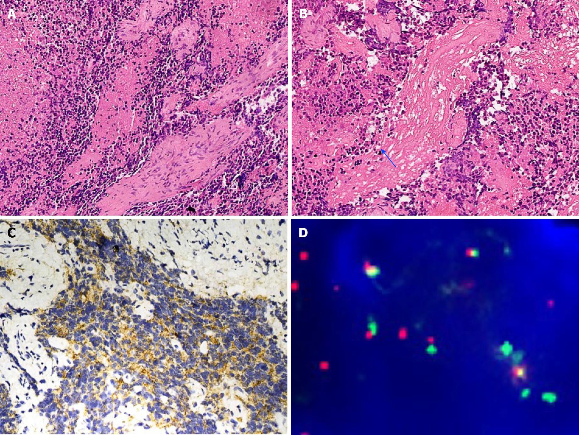
Figure 2 Pathological findings in case 1.
A: The poorly differentiated tumor showed a cluster appearance and a fibrovascular septum in case 1 (hematoxylin and eosin stain, × 200 magnification); B: Neuropil-like element in the tumor of case 1 (blue arrow, hematoxylin and eosin stain, × 200); C: The tumor cells were positive for synaptophysin in case 1 (× 200 magnification); D: The tumor cells show myelocytomatosis viral related oncogene, neuroblastoma derived amplification, which is indicated by the number of myelocytomatosis viral related oncogene, neuroblastoma derived signals (green) [more than 8 copies of the centrosomal probe signals (red)].
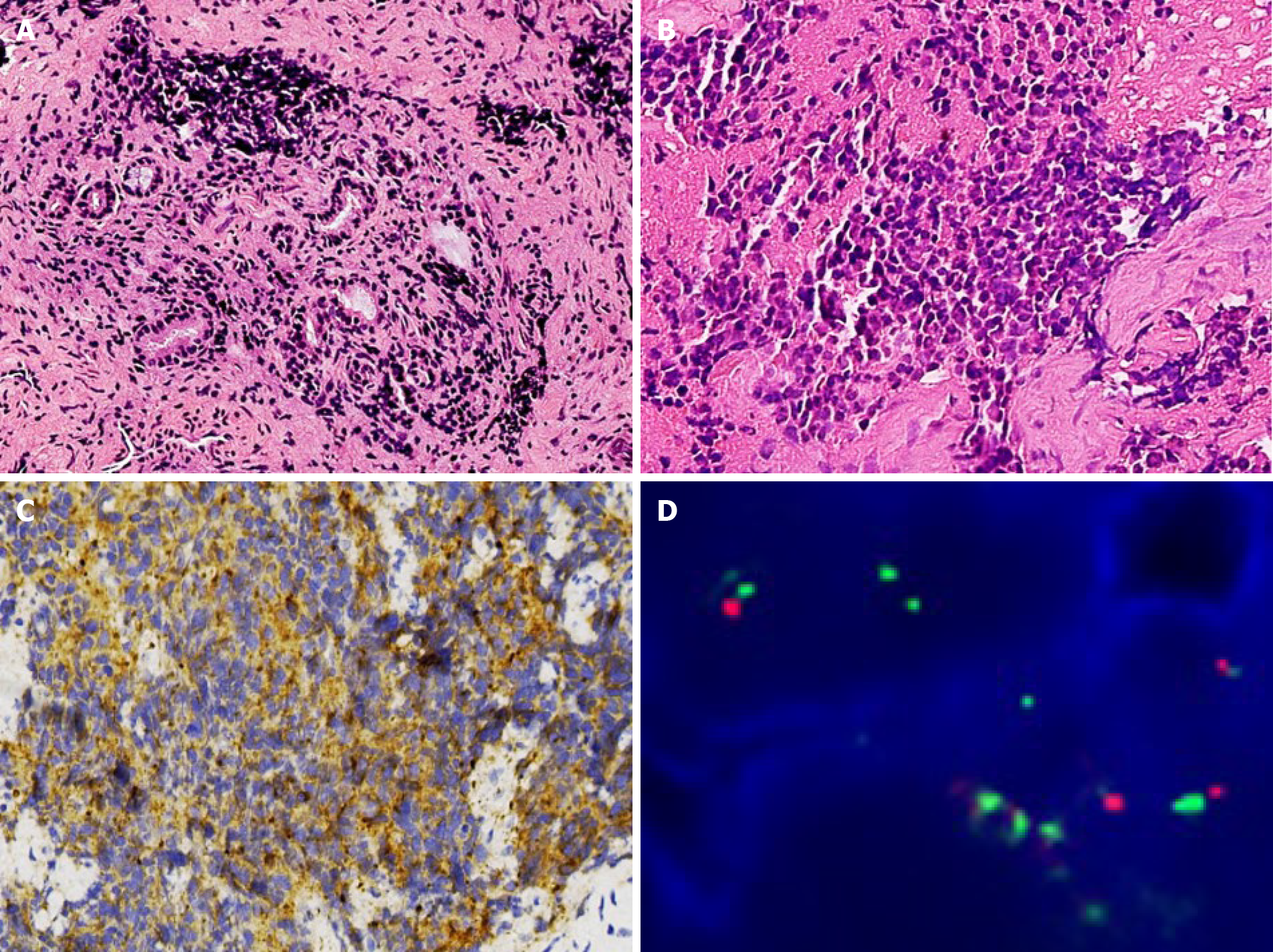
Figure 3 Pathological findings in case 2.
A: The poorly differentiated tumor also showed a cluster appearance in case 2 (hematoxylin and eosin stain, × 200 magnification); B: The tumor cells were round or oval and the nucleoli were not obvious in case 2 (hematoxylin and eosin stain, × 400 magnification); C: The tumor cells were positive for synaptophysin (× 200 magnification); D: The tumor cells showed myelocytomatosis viral related oncogene, neuroblastoma derived amplification, which is indicated by more myelocytomatosis viral related oncogene, neuroblastoma derived signals (green) [more than 8 copies of the centrosomal probe signals (red)].
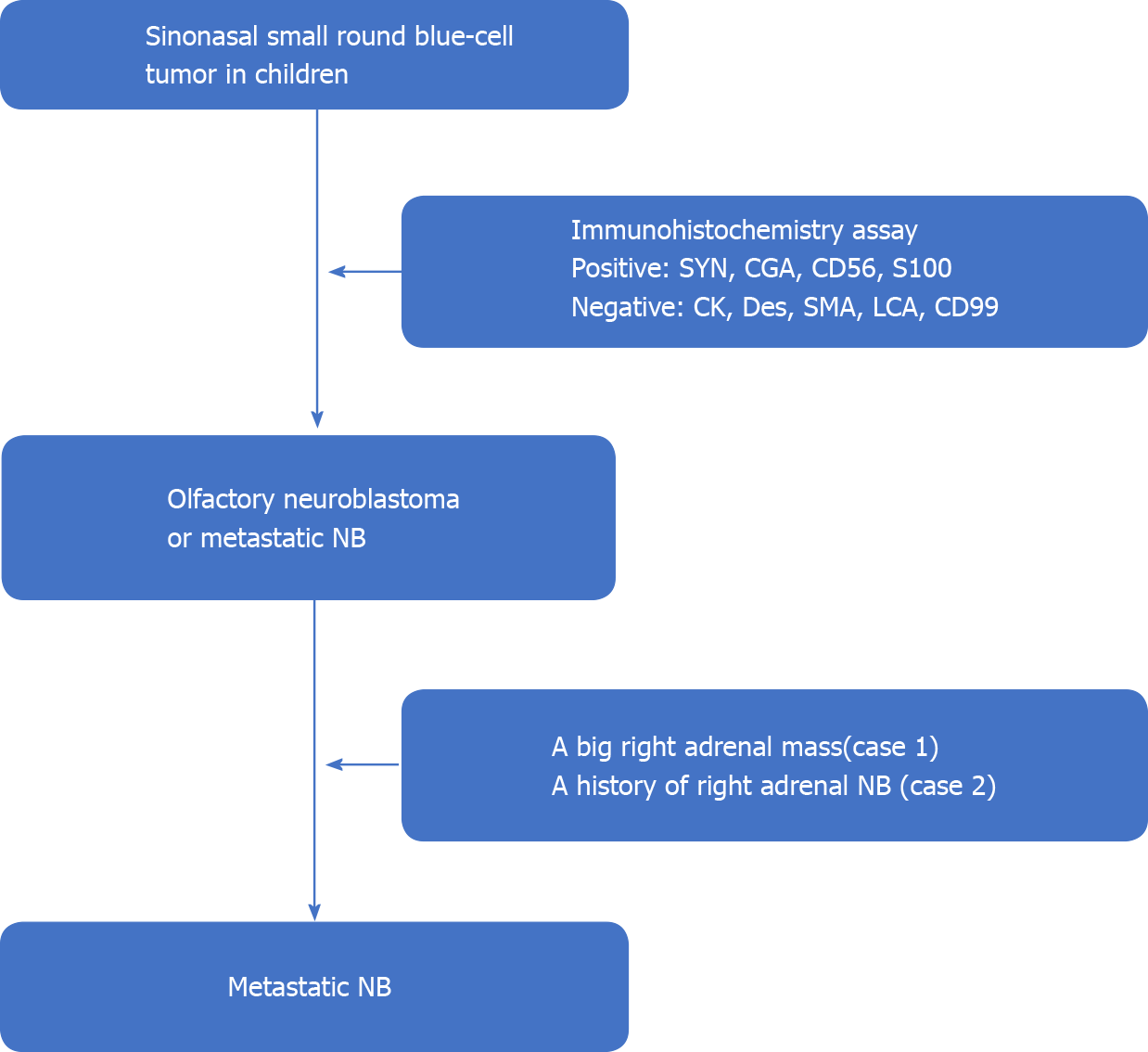
Figure 4 A schematic diagram of diagnosis procedure.
SYN: Synaptophysin; CGA: Chromogranin A; CD56: Cluster of differentiation 56; S100: S-100 protein; CK: Cytokeratin; DES: Desmin; SMA: Smooth muscle actin; LCA: Leucocyte common antigen; CD99: Cluster of differentiation 99; NB: Neuroblastoma.
Figure 5 Immunohistochemistry assay of paired like homeobox protein 2B.
A: Case 1; B: Case 2. The tumor cells were positive for paired like homeobox protein 2B in both cases. (× 400 magnification).
- Citation: Zhang Y, Guan WB, Wang RF, Yu WW, Jiang RQ, Liu Y, Wang LF, Wang J. Nasal metastases from neuroblastoma-a rare entity: Two case reports. World J Clin Cases 2021; 9(23): 6816-6823
- URL: https://www.wjgnet.com/2307-8960/full/v9/i23/6816.htm
- DOI: https://dx.doi.org/10.12998/wjcc.v9.i23.6816


